Figure 2. Morphology of photoreceptors isolated from dark- and light-adapted retinas. Cone cells were isolated from dark-adapted (A) and light-adapted retina (B). In both (A) and (B), ten typical cells are shown. Rods were isolated from dark-adapted (C) and light-adapted retina (D). Bars indicate 10 μm.
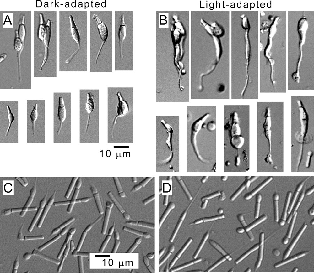
 Figure 2 of
Shimauchi-Matsukawa, Mol Vis 2008; 14:358-369.
Figure 2 of
Shimauchi-Matsukawa, Mol Vis 2008; 14:358-369.