Figure 1. Distribution pattern of opsin mRNA in carp photoreceptor cells. Antisense cRNA probes were hybridized with mRNA of rhodopsin
(A and B) and that of red-sensitive opsin (C and D) in dark-adapted (A and C) and light-adapted (B and D) retina. E and F: Controls were obtained using sense probes of red opsin in dark-adapted (E) and light-adapted (F) retina. Positive signals of rhodopsin in the outer segment layer in A and B are from the ellipsoid and the myoid of rods extending distally. Arrowheads indicate the approximate positions of the outer
limiting membrane. The following abbreviations were used: outer segment layer (OS), outer nuclear layer (ONL), and outer plexiform
layer (OPL). Bar indicates 20 μm in F.
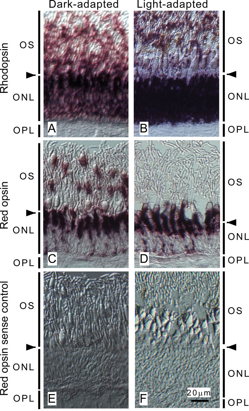
 Figure 1 of
Shimauchi-Matsukawa, Mol Vis 2008; 14:358-369.
Figure 1 of
Shimauchi-Matsukawa, Mol Vis 2008; 14:358-369.