Figure 4. VEGFR2 phosphorylation was decreased by injection of 50 ng VEGFab. Vascular endothelial growth factor receptor 2 (VEGFR2)
phosphorylation was noted mainly at the ganglion cell/nerve fiber layer and superficial and deep retinal vessels in noninjected
and IgG injected controls (2+, upper left). Phosphorylation of VEGFR2 was reduced after injection with 50 ng VEGFab (1+, lower
left). VEGFR1 phosphorylation was noted at the ganglion cell/nerve fiber layer and outer plexiform layer in noninjected and
IgG injected controls (2+, upper right). No change noted in 50 ng VEGFab-injected eyes (2+, lower right). All injections were
performed at p12 and eyes removed at p13. Green indicates phosphorylated VEGFR1 or phosphorylated VEGFR2; red indicates lectin-stained
vasculature. Central retina is toward the optic nerve, and peripheral retina is within vascularized retina near the junction
with avascular area.
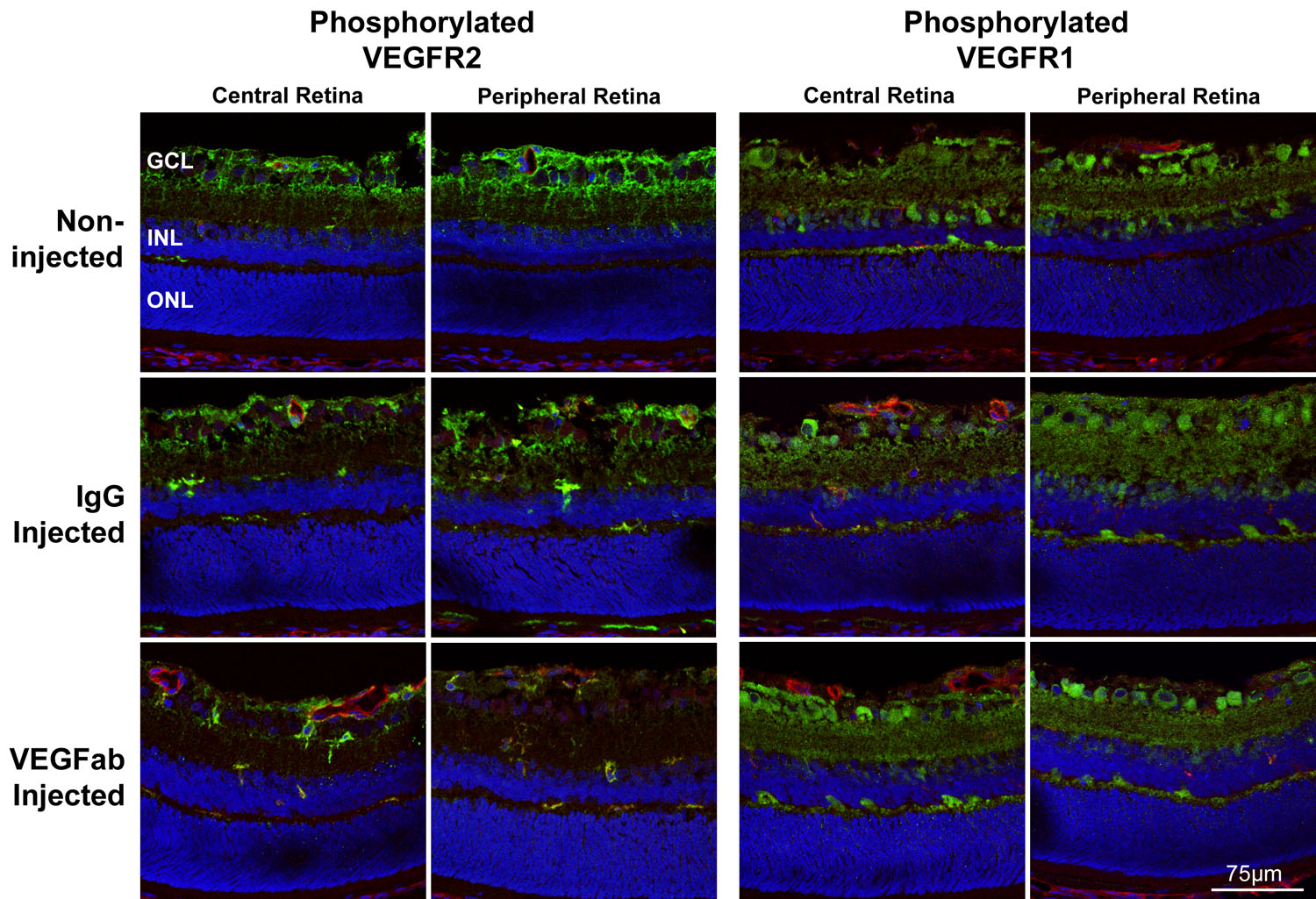
 Figure 4 of
Geisen, Mol Vis 2008; 14:345-357.
Figure 4 of
Geisen, Mol Vis 2008; 14:345-357.