Figure 3. Clock hours of intravitreous neovascularization and avascular/total retinal areas of lectin-stained retinal flat-mounts from
rat pups in 50/10 oxygen-induced retinopathy after intravitreous injection of neutralizing antibody to vascular endothelial
growth factor (VEGFab) or control nonimmune rat IgG. A: Mean clock hours of intravitreous neovascularization (IVNV)at p18
were significantly decreased by injection of either 25 ng or 50 ng VEGFab compared to IgG control (ANOVA p<0.001; * posthoc
Student’s t-tests p<0.001 compared to IgG). B: Mean clock hours of IVNV at p25 were significantly decreased by injection of 50 ng VEGFab
compared to IgG control (ANOVA p=0.003; * posthoc Student’s t-test p=0.003). C: Peripheral avascular/total retinal area was no different in retinas treated with either VEGFab dose compared
to IgG control at p18 (ANOVA p=0.238). D: At p25, the overall ANOVA for peripheral avascular/total retinal area was significant
(p=0.038). Posthoc testing showed the 50 ng dose of VEGFab was significantly decreased compared to the 25 ng dose (* posthoc
Student’s t-test, 25 ng versus 50 ng, p=0.038). However, neither dose of VEGFab was significantly different to control IgG.
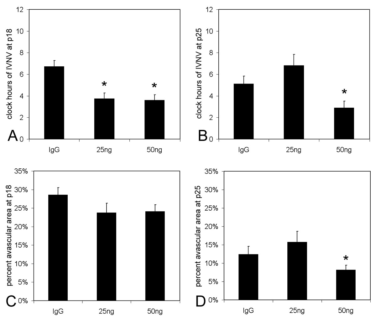
 Figure 3 of
Geisen, Mol Vis 2008; 14:345-357.
Figure 3 of
Geisen, Mol Vis 2008; 14:345-357.