Figure 3. Detection of TGF-β1 produced by splenocyte. Mice were sacrificed seven days after s.c. immunization. Spleens were removed
and grinded to prepare a single-cell suspension. These cells were incubated in a complete RPMI 1640 medium with OVA (100 μg/ml)
for 48 h in 24-well culture plates (2×10⁶ cells/well). Supernatants were collected for determining the quantity of TGF-β1 by ELISA. Data are the mean ± SEM (n=6 per
group). The asterisk indicates that p<0.05 as compared with the positive control.
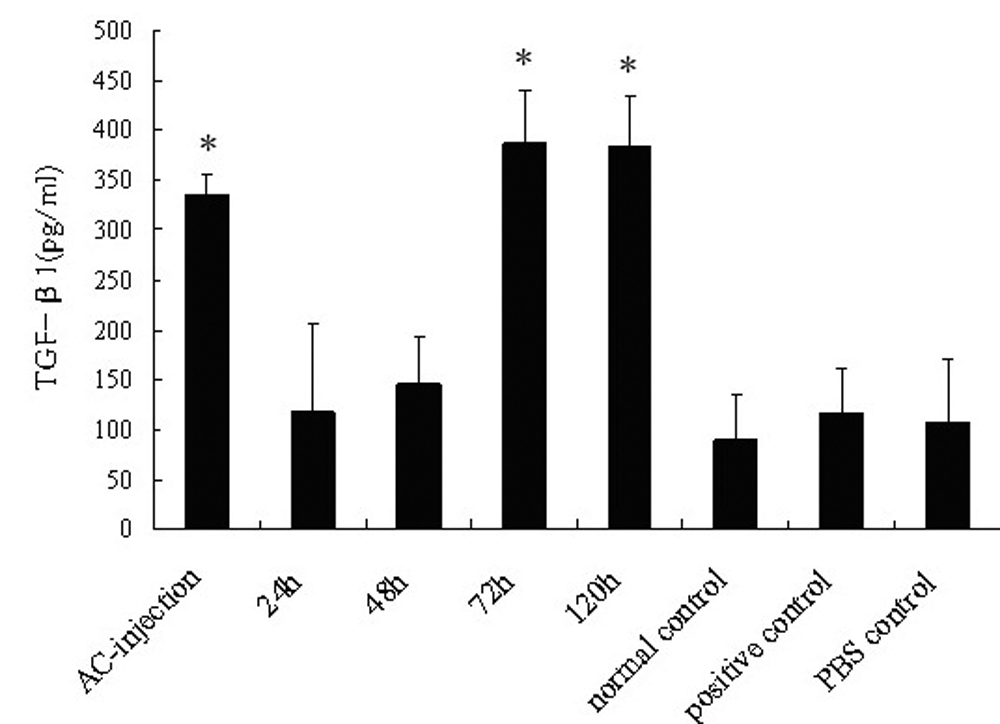
 Figure 3 of
Lei, Mol Vis 2008; 14:327-333.
Figure 3 of
Lei, Mol Vis 2008; 14:327-333.