Figure 2. Photograph (three-dimensional
lens) of a patient taken through a slit lamp. The lens opacity is
axial, extending from the anterior capsule to the posterior capsule. At
the anterior end, there is a round opacity about 2 mm in diameter,
placed slightly eccentrically toward 10 o'clock. There is a 1 mm x 1/2
mm blunt projection on the nasal side. On the temporal side, there
seems to be a fan-like opacity, which tends to hide the deeper
structure of the opacity, comprising about a dozen finger-like
projections going in all directions. The cataract appears like a
jellyfish.
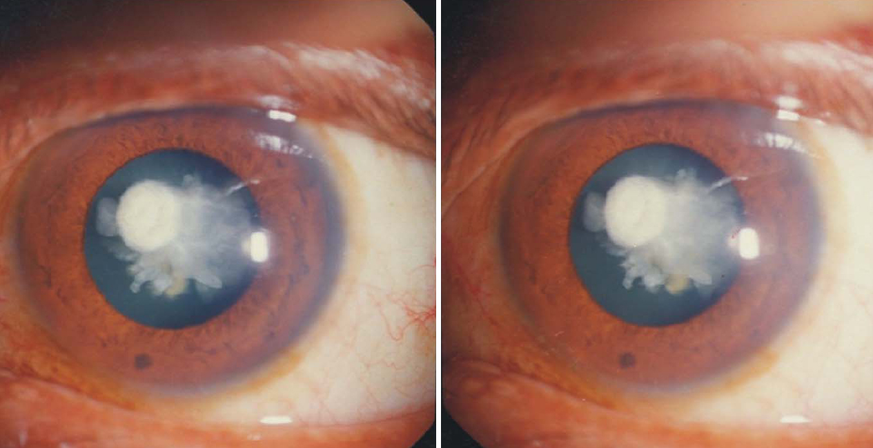
 Figure 2 of Vanita, Mol Vis 2008; 14:323-326.