Figure 3. Proliferation and matrix synthesis by keratocytes in spheroids. A: Cell cycle status of keratocytes in spheroids was visualized by incorporation of BrdU (green) in spheroids after three days
of culture in ADV medium containing FGF2 and PDGF. Cell nuclei were counterstained with propidium iodide (red). B: Keratan sulfate (green) and keratocan (red) were visualized after two weeks culture in ADV medium as described in Methods.
C: Lumican was stained in spheroids after two weeks culture in ADV medium as described in Methods. D: Collagen I was stained in spheroids after two weeks culture in ADV medium as described in Methods. E: Collagen V (green) and nuclear stain (red) were stained in spheroids after two weeks culture in ADV medium as described
in Methods. F: Collagen VI was stained in spheroids after two weeks culture in ADV medium as described in Methods. Bars show 50 μm.
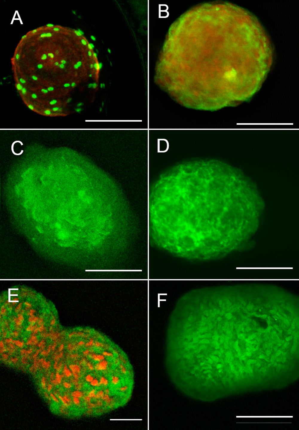
 Figure 3 of
Funderburgh, Mol Vis 2008; 14:308-317.
Figure 3 of
Funderburgh, Mol Vis 2008; 14:308-317.