Figure 2. All keratocytes participate in sphere formation. A: Freshly isolated primary keratocytes were cultured in polyHEMA-coated dishes either in Advanced-DMEM (ADV, dark solid bars)
or in Advanced-DMEM containing transforming growth factor beta-1 (TGFβ-1), fibroblast growth factor 2 (FGF2), and platelet
derived growth factor BB (PDGF; light-stippled bars). Viable cells were labeled with Calcein AM, and the number labeled cells
was determined by direct counting after trypsinization. Cell number is shown as a percentage of viable cells added to the
original culture. B: Primary stromal cells were plated in Advanced-DMEM containing 10 ng/ml FGF2 under attachment conditions (solid lines) or
as spheroids in polyHEMA-coated dishes (broken lines). Cells numbers were estimated by conversion of the dye Alamar Blue to
a fluorescent form and normalized to the number of cells added to the culture. Error bars show the SD of triplicate analyses.
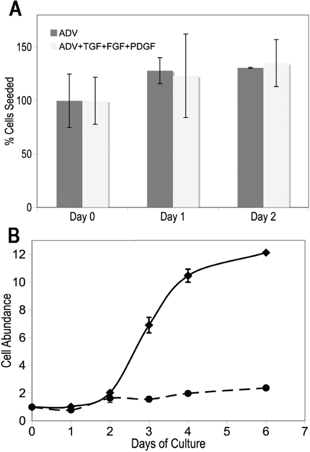
 Figure 2 of
Funderburgh, Mol Vis 2008; 14:308-317.
Figure 2 of
Funderburgh, Mol Vis 2008; 14:308-317.