Figure 1. Spheroid formation by primary keratocytes. Freshly isolated primary bovine keratocytes were cultured in serum-free conditions
on standard tissue culture plastic (A) or in vessels coated with polyHEMA, which prevented cell attachment (B,C,D). In two days (B), cells had formed aggregates, which condensed into smooth spheroids after two weeks of culture (C). D: In this panel, two-week spheroids were stained with vital dye Calcein AM to detect live cells (green) and propidium iodide
(red) to detect dead cells. Scale bars show 50 µm.
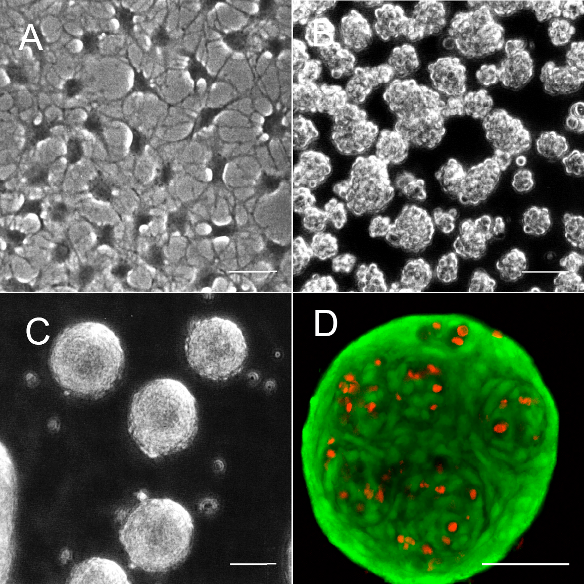
 Figure 1 of
Funderburgh, Mol Vis 2008; 14:308-317.
Figure 1 of
Funderburgh, Mol Vis 2008; 14:308-317.