Figure 5. Confocal images of AQP4 labeling in the retina after 48 hours of occlusion. Composite confocal micrograph of a transverse
section of the retina after 48 h of occluder wear is displayed. The left section demonstrates the differential interference
contrast image and the right section illustrates the AQP4 immunofluorescent image in red. The middle section shows both images
superimposed. Note that AQP4 labeling is predominant in the nerve fiber, ganglion cell and inner plexiform layers of the inner
retina, just as demonstrated by the normal eye (see Figure 1). However, the intensity of labeling is greater along the vitreal
border in the form-deprived eye than in a normal eye. Bar is 50 μm.
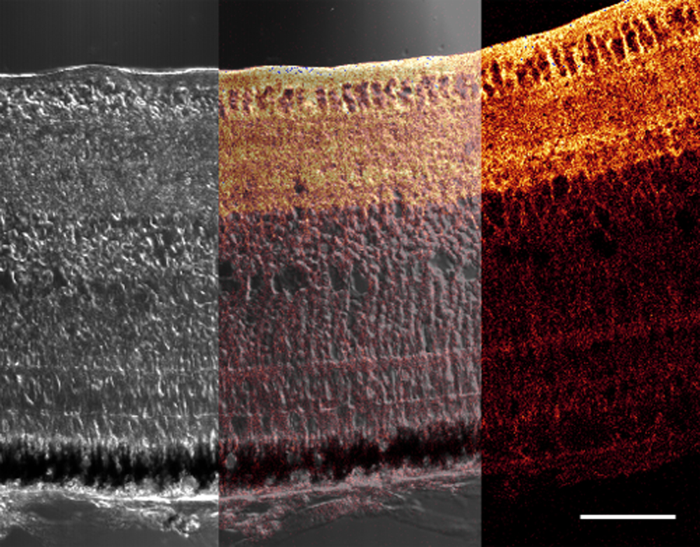
 Figure 5 of
Goodyear, Mol Vis 2008; 14:298-307.
Figure 5 of
Goodyear, Mol Vis 2008; 14:298-307.