Figure 4. Intensity of AQP4 antibody
labeling in the nerve fiber layer of chick. The mean relative
fluorescence (on an arbitrary scale of 0 to 255) of AQP4/Alexa 596
conjugated antibodies expressed in the nerve fiber layer of the retinae
of age-matched normal control eyes (triangles) and both non-deprived
eyes (squares) and fellow eyes that experienced form deprivation
(diamonds) is plotted against time from initiation of occlusion of the
experimental eye. Note that initially intensity of AQP4 labeling is
slightly raised in normal eyes and falls with time and increase in eye
size. In contrast, occlusion initially causes significant elevation of
labeling intensity particularly in the form-deprived eye, but also in
the non-occluded fellow eye, which falls with duration of
form-deprivation to almost reach control levels after 4 days. Error
bars are 1 standard error.
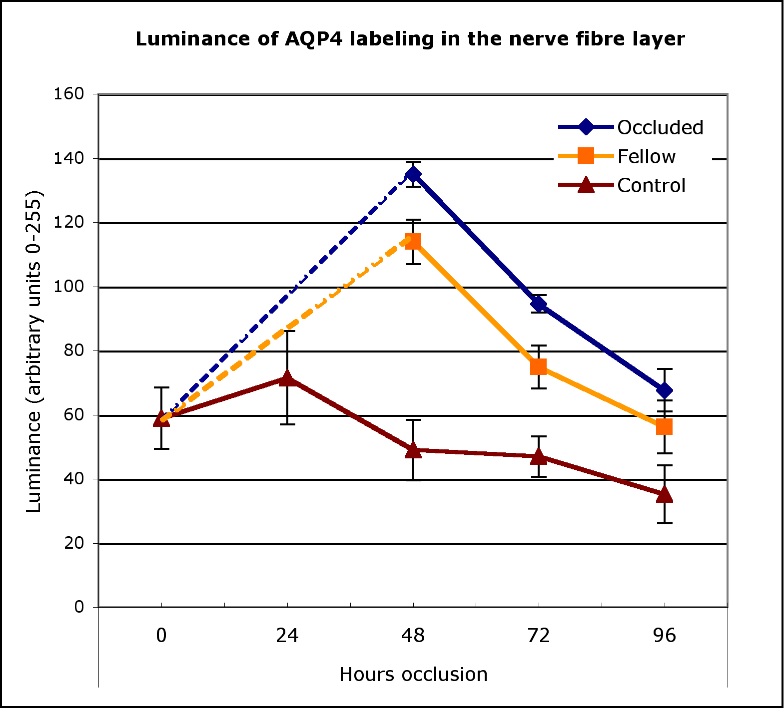
 Figure 4 of Goodyear, Mol Vis 2008; 14:298-307.