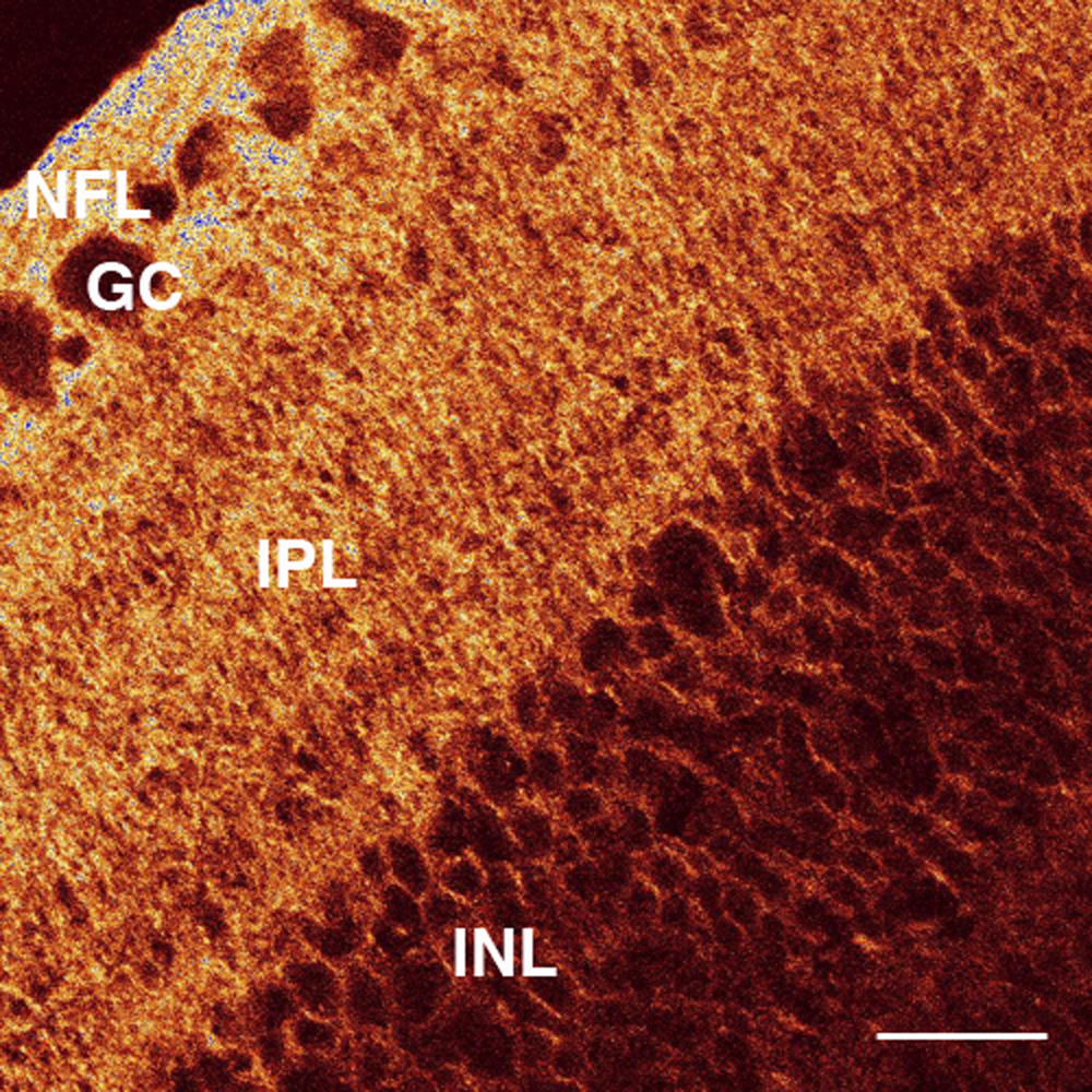
Figure 3. AQP4 labeling of the chick
retina. A confocal micrograph of a transverse section of the retina
from a form-deprived eye after 72 h of occluder wear is shown. There is
strong labeling in the nerve fiber layer (NFL), ganglion cell (GC)
layer, and inner plexiform layer (IPL) but very weak labeling in the
inner nuclear layer (INL). Note the branching brush-like appearance of
the pattern of AQP4 expression and also the appearance of several
sublaminae in the IPL, which is similar to the previously described
morphology of the Müller cell [
51]. Bar is 20 µm.
 Figure 3 of Goodyear, Mol Vis 2008; 14:298-307.