Figure 1. AQP4 immunolabeling of the normal chick retina plus negative controls. A: Confocal micrograph (40X magnification) with red AQP4 immunolabeling (to the right) of a transverse section of a normal chick
retina merged (in middle) with the accompanying differential interference contrast image (to the left) is shown. The layers
of the retina are indicated: NFL nerve fiber layer, GC ganglion cell layer, IPL inner plexiform layer, INL inner nuclear layer,
IS inner segments of photoreceptors, OS outer segments of photoreceptors, RPE retinal pigment epithelium. It can be seen that
labeling is most intense within the NFL, GC and IPL layers. Only very weak label is visible in the INL and photoreceptor region.
B: Negative control with the primary antibody omitted indicates very weak red non-specific labeling (to the right) merged with
the accompanying differential interference contrast image (to the left). C: Tissue pre-incubated with purified peptide before AQP4 labeling is shown indicating that blocking of AQP4 sites with the
purified peptide successfully prevents binding of the introduced AQP4 antibodies and apparent red immunolabel. Bars=50 μm.
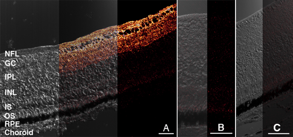
 Figure 1 of
Goodyear, Mol Vis 2008; 14:298-307.
Figure 1 of
Goodyear, Mol Vis 2008; 14:298-307.