Figure 6. The presence of free DNA and ROS in the x-ray cataract. Using LSCM with fluorescent dyes for DNA and ROS, integrated sequential
sections from the surface down to 120 μm beneath the lens surface revealed the following in a 3.5+ x-ray cataract that developed
5 months post-radiation: A: Free and particulate DNA is present in the left upper part of the irradiated lens. B: A control animal at the same age and time is free of the abnormal internal DNA content. C: A great amount of internal ROS is present in the irradiated lens at several depths. D: The control animal’s lens shows a very small amount of ROS near the lens surface. The dotted lines were placed on the lens
sections to demarcate the position of the lens surface.
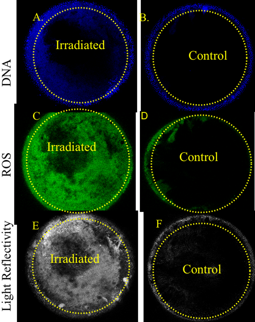
 Figure 6 of
Wolf, Mol Vis 2008; 14:274-285.
Figure 6 of
Wolf, Mol Vis 2008; 14:274-285.