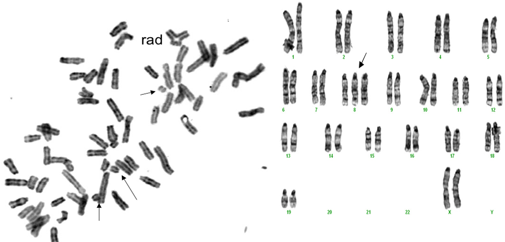
Figure 10. Chromosomal aberrations are present in LEC cultured for 10 days after removal 72 h post-irradiation. Left panel shows LEC
from an irradiated mouse taken 72 h after irradiation and cultured for a total of 12 days in 3% O
2, treated with colchicine for 12 h to arrest metaphases and then fixed as described in Methods, and stained with H&E. After
this procedure, the metaphase spreads were read for chromosomal abnormalities. Disarray of chromosomes and broken chromosomes
in the cell from the x-irradiated animal made the selection of chromosomal pairings impossible. Arrows point to broken chromosomes,
and an adherence is seen at the uppermost portion in this near-tetraploid set. On the right is a spread from a same age non-irradiated
mouse. One abnormality, a trisomy, is noted by the arrow. For comparisons of the damages in the several cells studied in each
group see
Table 1. Magnification 400X.
 Figure 10 of
Wolf, Mol Vis 2008; 14:274-285.
Figure 10 of
Wolf, Mol Vis 2008; 14:274-285.