Figure 2. Funduscopic photograph of a patient with Stargardt disease. Funduscopy revealed a maculopathy with retinal pigment epithelium
atrophy and hyperpigmentation and few central yellowish flecks. A slight temporal papillary pallor was present and retinal
vessels showed no constriction.
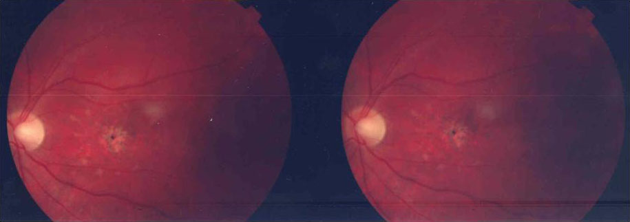
 Figure 2 of
Riveiro-Alvarez, Mol Vis 2008; 14:262-267.
Figure 2 of
Riveiro-Alvarez, Mol Vis 2008; 14:262-267.