Figure 4. In vitro expression of c cyclic guanosine monophosphate (cGMP) in the nerve fiber layer and inner nuclear layer of a healthy
or detached retina. Small pieces of retina were incubated with IBMX (a non-specific PDE inhibitor) and the soluble guanylyl
cyclase (sGC) stimulator sodium nitroprusside (SNP). The expression of cyclic GMP (cGMP: green color) in the retina was analyzed
by immunohistochemistry and nuclei were counterstained with Hoechst 33342 (red). Panels A and B show a similar cGMP expression
in nerve fiber layer of the normal (A) or detached (B) retina. Panel C (normal retina) and Panel D (detached retina) show
cGMP staining in the inner nuclear cells stained in red. Scale bar represents 50 μm.
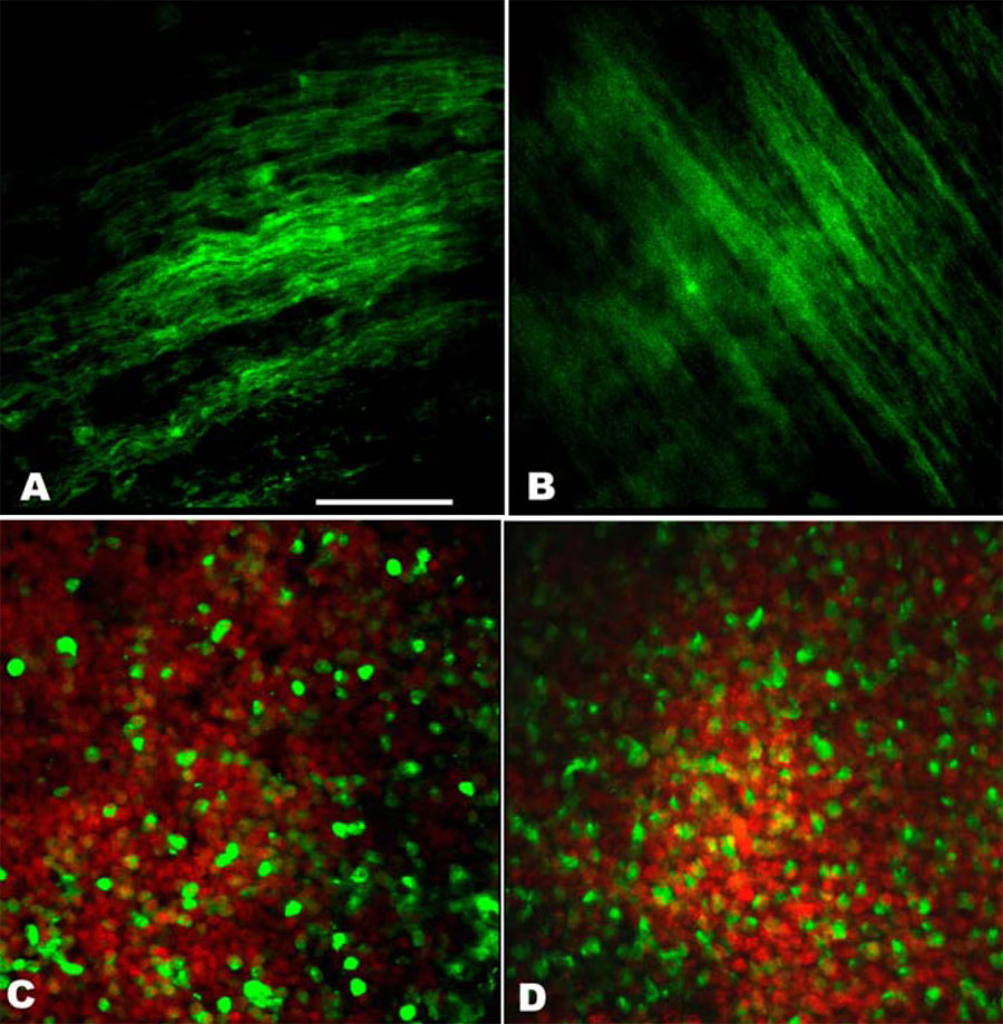
 Figure 4 of
Diederen, Mol Vis 2008; 14:255-261.
Figure 4 of
Diederen, Mol Vis 2008; 14:255-261.