Figure 3. In vitro expression of cyclic guanosine monophosphate (cGMP) in the outer plexiform layer of a detached retina. Small pieces
of retina were incubated with IBMX (a non-specific PDE inhibitor) and the particulate guanylyl cyclase (pGC) stimulator atrial
natriuretic peptide (ANP). The expression of cGMP (cGMP: green color) in the retina was then analyzed by immunohistochemistry
and nuclei were counterstained with Hoechst 33342 (red). Following stimulation with ANP in the presence of IBMX, both the
detached and healthy retina showed cGMP-immunolabeling in the outer plexiform layer. Panel A shows the labeling of the outer
plexiform layer of a detached retina with Hoechst. Panel B shows the cGMP stain (green) of the outer plexiform layer probably
representing photoreceptor axons and dendrites. Panel C is a double staining exposure of the nuclei and cGMP. Scale bar represents
20 μm.
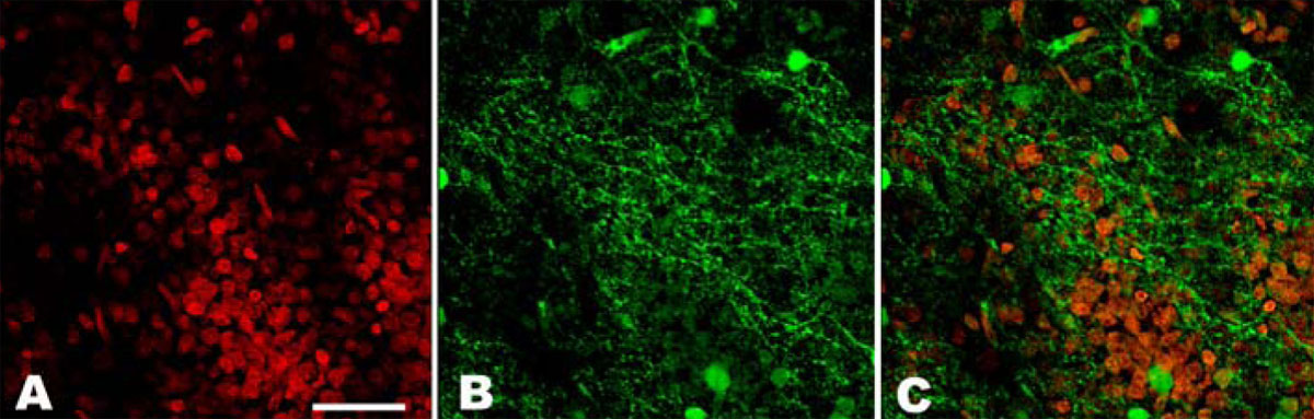
 Figure 3 of
Diederen, Mol Vis 2008; 14:255-261.
Figure 3 of
Diederen, Mol Vis 2008; 14:255-261.