Figure 2. In vitro expression of cyclic guanosine monophosphate (cGMP) in the photoreceptor layer of a normal or detached pig retina.
Small pieces of retina were incubated with IBMX (a non-specific PDE inhibitor) and the particulate guanylyl cyclase (pGC)
stimulator atrial natriuretic peptide (ANP). The expression of cGMP (cGMP: green color) in the retina was then analyzed by
immunohistochemistry and nuclei were counterstained with Hoechst 33342 (red). Under these conditions no cGMP signal was visible
in the photoreceptor layer of the healthy retina (Panel A). The detached retina on the other hand displayed strong cGMP expression
in the photoreceptor layer (Panels B and C). Panel B is a double staining exposure for the nuclei and cGMP, showing the presence
of cGMP in the nuclei of the cells (yellow) and in the cytoplasma (green). In Panel C we only show the green signal, showing
strong cGMP immunoreactivity in the same piece of retina. Scale bar represents 25 μm.
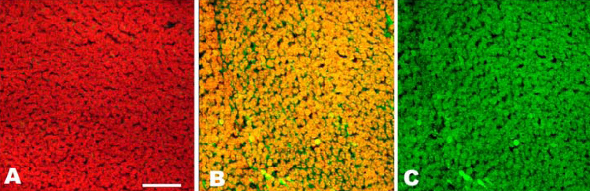
 Figure 2 of
Diederen, Mol Vis 2008; 14:255-261.
Figure 2 of
Diederen, Mol Vis 2008; 14:255-261.