![]() Figure 6 of
Gu, Mol Vis 2008;
14:20-28.
Figure 6 of
Gu, Mol Vis 2008;
14:20-28.
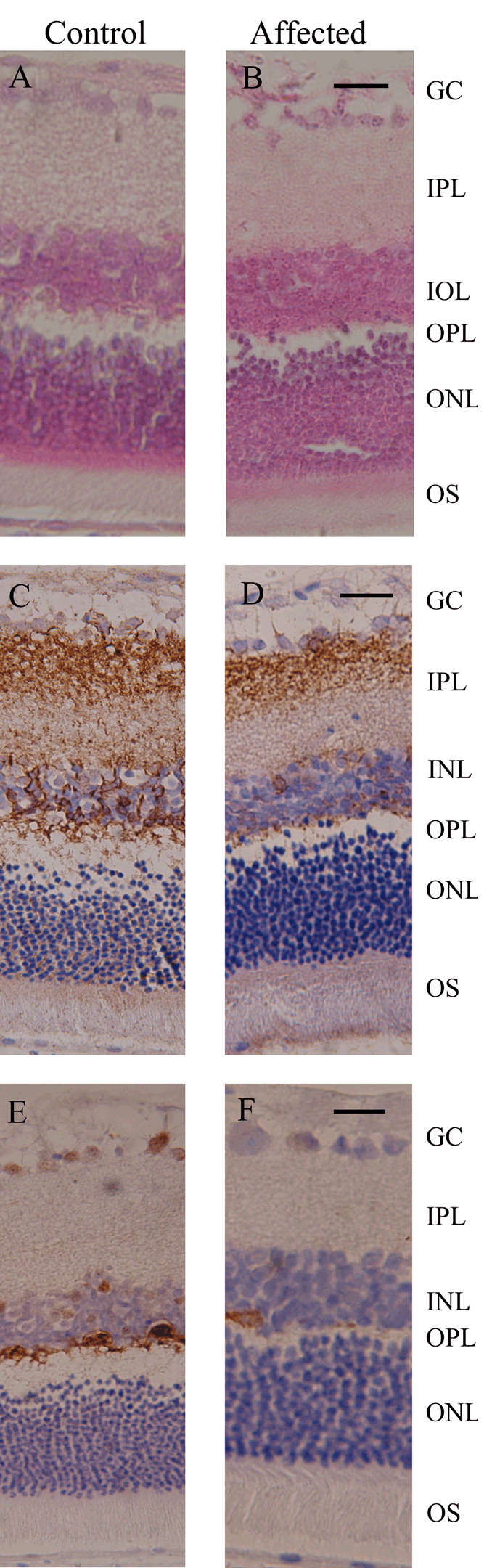
Figure 6. Histology and immunolabeling of retinal sections from control and affected rats
Retinal sections of control and affected rats were stained with hematoxylin and eosin (A and B). Bioplar and horizontal cells were identified with antibodies directed to PKCα (C and D) and calbidin (E and F). The following abbreviations are used: ganglion cells layer (GC), inner plexiform layer (IPL), inner nuclear layer (INL), outer plexiform layer (OPL), outer nuclear layer (ONL), and outer segment (OS). The scale bar represents 20 μm.