Figure 8. IKKΒ-siRNA inhibits the expression of IKKβ on both the mRNA and protein level. A: mRNA transcription of IKKβ in HTFs assessed by real-time RT-PCR 24 h after 5-100 nM IKKΒ-siRNA was transfected. The normalized IKKβ mRNA level of non-transfected HTFs is taken as 1.0 (the asterisk indicates a p<0.05, mean±SD, n=3). B: Protein levels of IKKβ demonstrated by western blot. C-N: Confocal laser scanning microscopy images shows the intracellular distribution of NF-κB in HTFs. Green fluorescence indicates
the intracellular distribution of phosphated NF-κB, and blue fluorescence represents the DAPI counterstained cell nuclei.
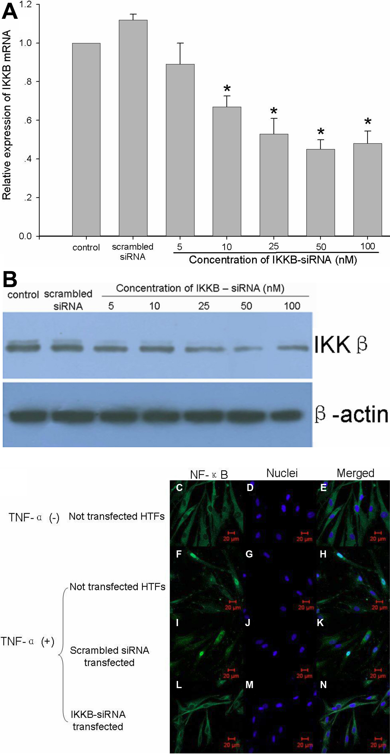
 Figure 8 of
Duan, Mol Vis 2008; 14:2616-2628.
Figure 8 of
Duan, Mol Vis 2008; 14:2616-2628.