Figure 8. mPins protein localization in
the adult mouse cornea and ciliary body by immunohistochemistry. A
and C show localization of the mPins protein in the adult
cornea and ciliary body. B and D are the corresponding
confocal views. In the cornea, mPins protein is observed in the
epithelium (CEp), stroma (CS), and endothelium (CEn; A). The
confocal view (B) shows mPins localization in the stromal
keratocytes (ke, arrows). In the ciliary body, mPins protein is
detected in the nonpigmented ciliary epithelial cells (NPCE). No
significant labeling is observed in the pigmented ciliary epithelial
(PCE) cells (C and D).
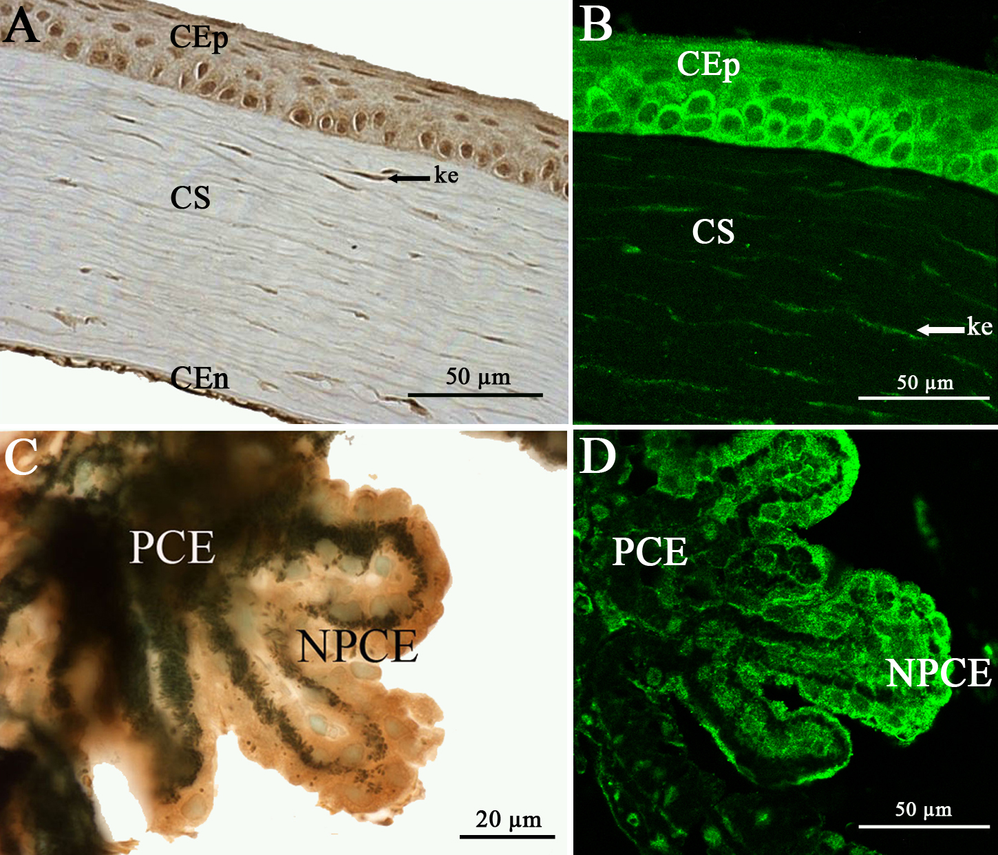
 Figure 8 of Raji, Mol Vis 2008; 14:2575-2596.