Figure 7. mPins protein localization in
the adult mouse retina by immunohistochemistry. A-C show
mPins protein localization by DAB chromogen immunochemistry. A
shows the DAB-immunonegative control. D-F show confocal
views of mPins protein localization by immunofluorescence; E
shows the immunonegative control. mPins protein is detected in all
retinal nuclear layers: the ganglion cell layer (GCL), the inner
nuclear layer (INL), and the outer nuclear layer (ONL). The protein is
also observed in the inner plexiform layer (IPL), with weak labeling
detectable in the outer plexiform layer (OPL; B and C).
mPins protein is also observed in the retinal pigment epithelium (RPE)
and in the choroidal melanocytes (Ch; C). Higher magnification
also shows strong immunolabeling in the inner segments of the
photoreceptors (IS; C). Moreover, in the INL, mPins labeling is
stronger on either side of the layer than in its center (C,
asterisks). Confocal views confirm this distribution of protein and
show the mPins protein to be present principally in the cellular
membrane of the INL and ONL cells (D and F).
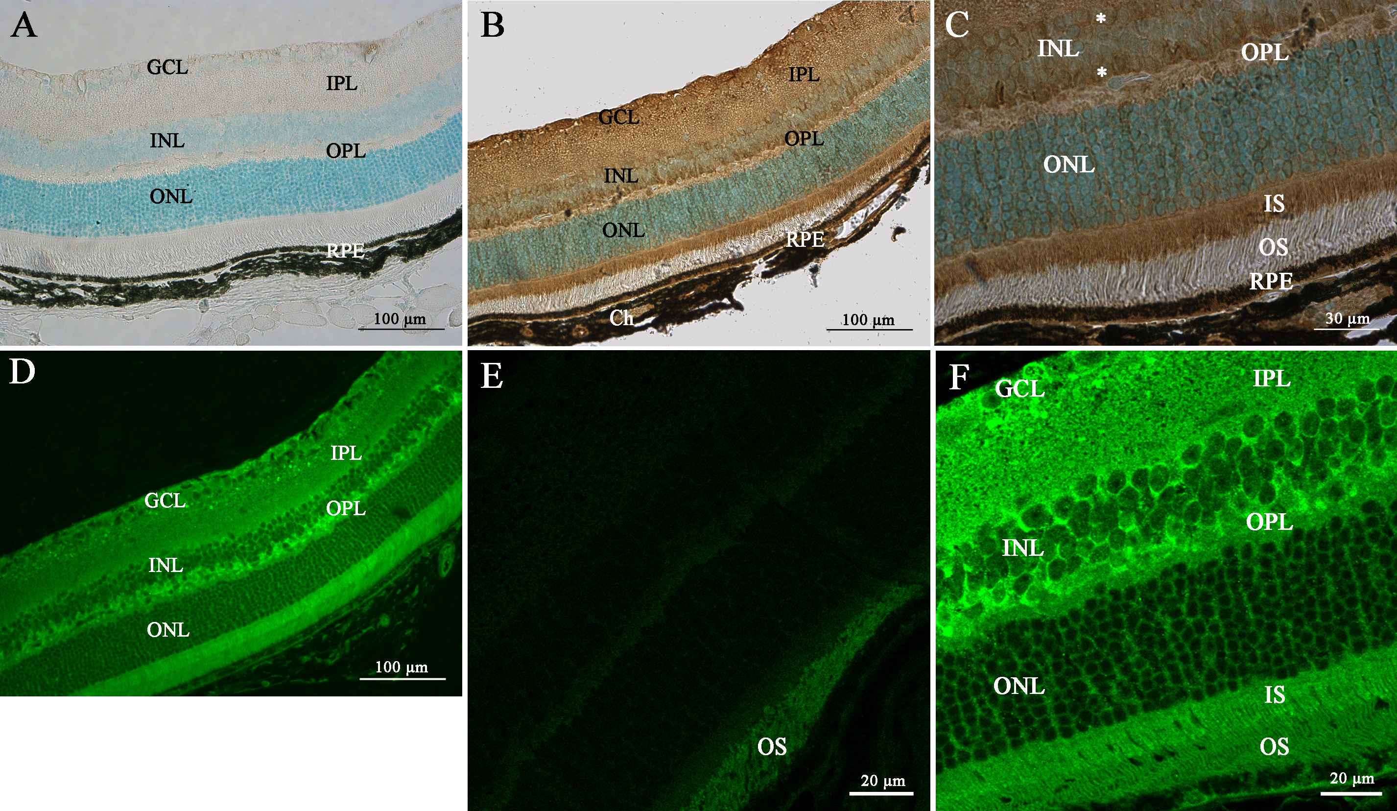
 Figure 7 of Raji, Mol Vis 2008; 14:2575-2596.