Figure 6. mPins protein localization in
postnatal mouse retina by immunohistochemistry. mPins protein
localization is shown in the developing mouse retina at postnatal
stages P3 (B), P5 (C), and P12 (D). At stages P3 (B)
and P5 (C), mPins protein is detected primarily in the ganglion
cell layer (GCL) and the neuroblastic layer (NbL). In the retina at
stage P12, mPins is observed in the GCL, the inner nuclear layer (INL),
and the outer nuclear layer (ONL; D). Moreover, in the INL,
mPins labeling was found to be more intense on either side of this
layer than in its center (asterisks). Immunolabeling is also detected
in the inner plexiform layer (IPL), but no labeling is observed in the
outer plexiform layer (OPL) or in the photoreceptor segments (PS; D).
A shows Diaminobenzidine (DAB) immunonegative control.
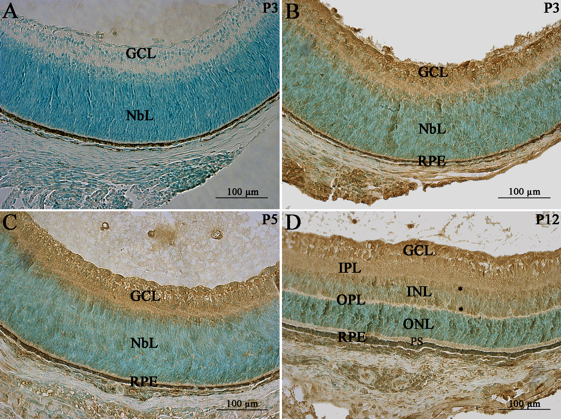
 Figure 6 of Raji, Mol Vis 2008; 14:2575-2596.