Figure 5. mPins protein localization in
embryonic mouse retina by immunohistochemistry. mPins protein
localization is shown in embryonic mouse retina at stages E12.5 (A),
E14.5 (B), E16.5 (D), and E18.5 (E and F).
mPins protein distribution is also shown in lens at E14.5 (C)
and E18.5 (G). Orientation of mouse eye sections is specified
below the figure. From E12.5 to E18.5, mPins protein is detected in
neuroblastic layer (NbL; A, B, D, E,
and F). Furthermore, mPins immunolabeling is stronger in the
inner part of the NbL from E16.5 (D-F). At this stage, the mPins
protein is also observed in the ganglion cell layer (GCL; F).
The mPins protein is also found in the lens with a specific
distribution depending on the stage (C and G). At the
E14.5 stage (C), immunostaining is observed in the anterior
region, whereas staining is observed only in the posterior region of
the lens by E18.5 (G). Moreover, mPins protein is observed in
the corneal epithelium, the presumptive cornea from E12.5 (A and
E). H shows Diaminobenzidine (DAB) immunonegative
control.
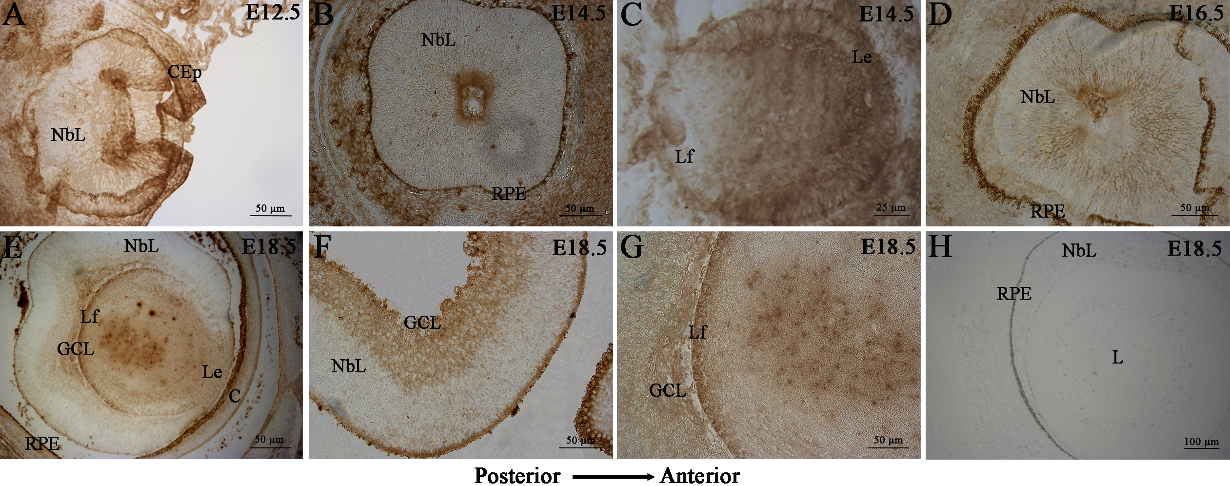
 Figure 5 of Raji, Mol Vis 2008; 14:2575-2596.