Figure 3. mPins mRNA localization
in the postnatal and adult retina by in situ PCR hybridization. mPins
labeling is shown in the postnatal retina at P3 (A) and P12 (B),
and in the adult retina (D and E). C and F
are higher magnifications of B and E, respectively. G
and H show negative controls at P3 and in adult retina,
respectively, using the mPins sense probe. At the P3 stage, significant
mPins mRNA labeling is detected in the neuroblastic (NbL) and
ganglion cell layers (GCL; A). In the retina, at the P12 stage,
significant mPins mRNA is detected in the GCL, the inner
nuclear layer (INL), the outer nuclear layer (ONL), and in the inner
segments of the photoreceptors (IS; B). High magnification of
retina at this stage shows that mPins mRNA labeling is more
intense on either side of the INL (C, asterisks). No significant
labeling is observed in either of the plexiform layers (B and C).
A similar pattern of mPins expression is observed in the adult
retina (D and E). mPins mRNA is detected in cell
bodies on sections of the optic nerve (ON; E). Significant mPins
labeling is also detected at this stage in the retinal pigment
epithelium (RPE) and choroidal melanocytes (Ch; F).
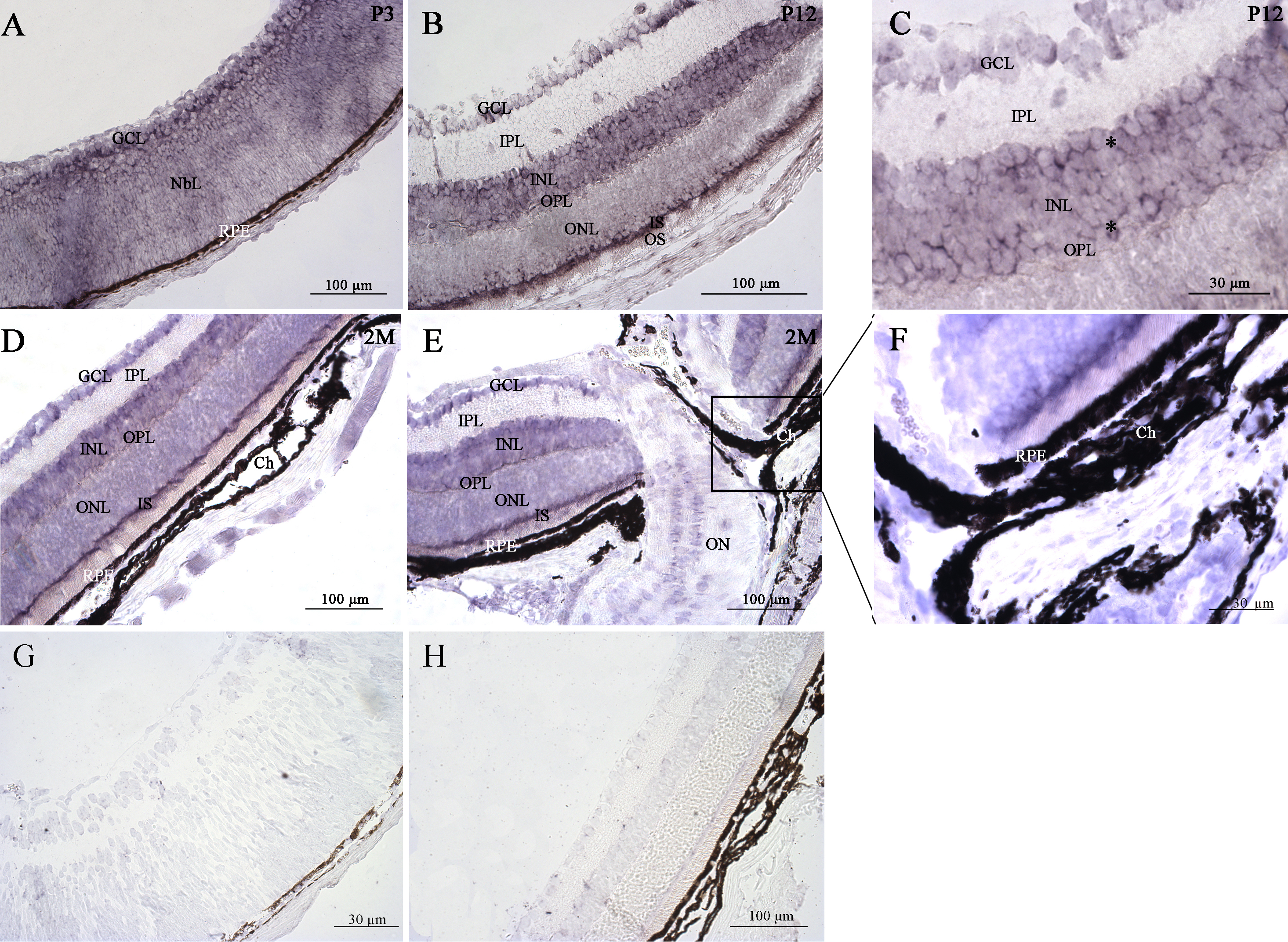
 Figure 3 of Raji, Mol Vis 2008; 14:2575-2596.