Figure 2. mPins mRNA localization
in mouse embryo by radioactive in situ hybridization. mPins
labeling with dark-field illumination (left) and the corresponding
bright-field (right) are shown for stages E11.5 (A), E12.5 (B),
E13.5 (C), E14.5 (D), E15.5 (E), E16.5 (F),
E17.5 (G) and E18.5 (H and I). Dark-field
negative controls using sense (J, left) and scramble (J,
right) are also shown. mPins transcripts are detected in the
neuroblastic layer (NbL) of the retina from E11.5 to E18.5 of embryonic
development (A-I). The corresponding bright-field views
confirm these observations (black grains). At E18.5, no significant
signal is detected in the ganglion cell layer (H). At the same
stages, we also observe a strong signal at the posterior face of the
lens, corresponding to secondary fiber cells (I, asterisk).
Negative controls using sense (J, left) and scramble (J,
right) probes gave no significant signal.
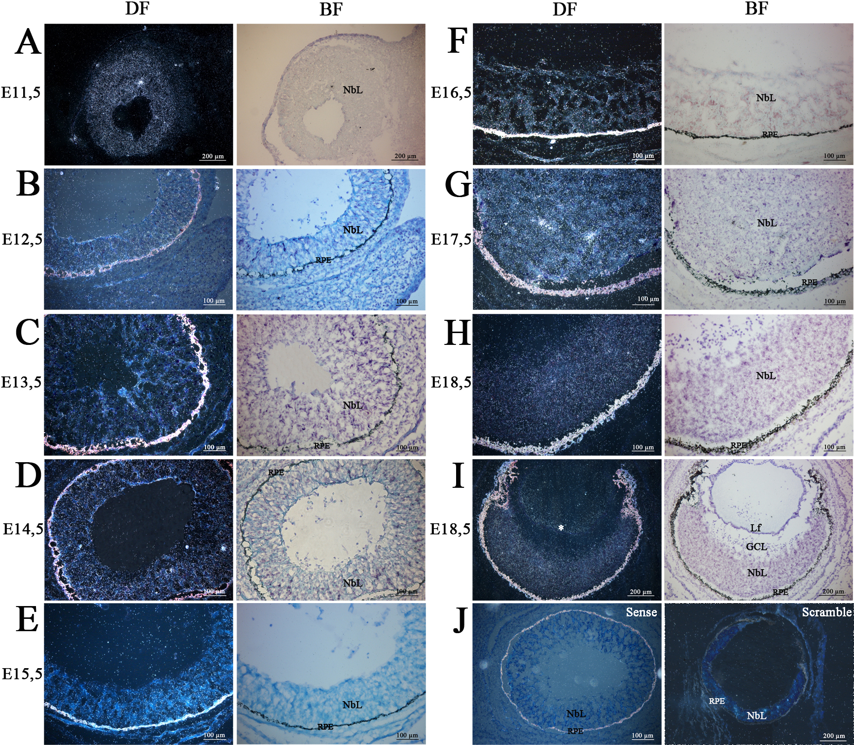
 Figure 2 of Raji, Mol Vis 2008; 14:2575-2596.