Figure 8. TSA induced the senescence-like state of corneal fibroblasts as indicated by β-galactosidase staining. TSA at 400 nM induced
reduced saturation densities in corneal fibroblasts and more prominent β-galactosidase activity (B) than the untreated cells of equal density (A), while the positive control cells (treated with 100µM H2O2) showed enhanced staining of β-galactosidase (C). D: Image quantitative analysis of different β-galactosidase staining densities; the number of asterisks represents different
densities with the three astrisks indicating the strongest staining of β-galactosidase.
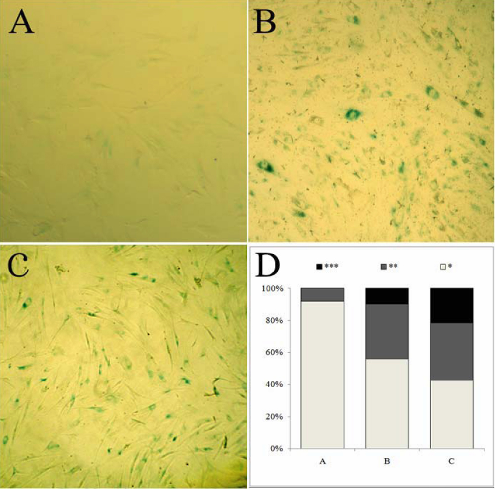
 Figure 8 of
Zhou, Mol Vis 2008; 14:2556-2565.
Figure 8 of
Zhou, Mol Vis 2008; 14:2556-2565.