Figure 6. Determination of molecular mass
by dynamic light scattering method of UV-A-unexposed and exposed WT
αB-crystallin and its three deamidated mutants. A multiage laser light
scattering instrument (Wyatt Technology, Santa Barbara, CA) coupled to
a HPLC was used to determine the molecular mass of the WT protein and
its deamidated mutant proteins. The figure shows elution profiles at
280 nm of UV-A-unexposed (____) and UV-A exposed (-----)
αB-Asn146Asp mutant protein from a TSK G-5000PWXL column as
well as the molecular mass. Similar to the UV-A-exposed αB-Asn146Asp
mutant protein profile, a high molecular weight (HMW) protein peak that
eluted first (at about 10 min) followed by a crystallin peak (at about
15 min) were also observed for WT αB-crystallin and the αB-Asn78Asp and
αB-Asn78/146Asp mutant proteins.
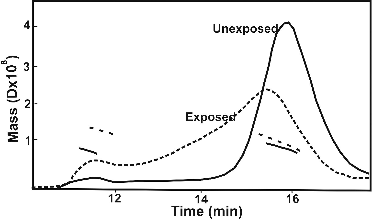
 Figure 6 of Mafia, Mol Vis 2008; 14:234-248.