Figure 1. SDS–PAGE analysis of UV-A-exposed and unexposed WT αB-crystallin and its three deamidated mutant species. After UV-A-exposure
of varying doses (0, 20, and 50 J/cm2, shown at the bottom of the gel), the WT αB-crystallin and its three deamidated mutant proteins (αB-Asn78Asp, αB-Asn146Asp,
and αB-Asn78/146Asp) were analyzed. Increased dimerization of each protein and degradation, particularly in the deamidated
species, were observed following UV-A-exposure.
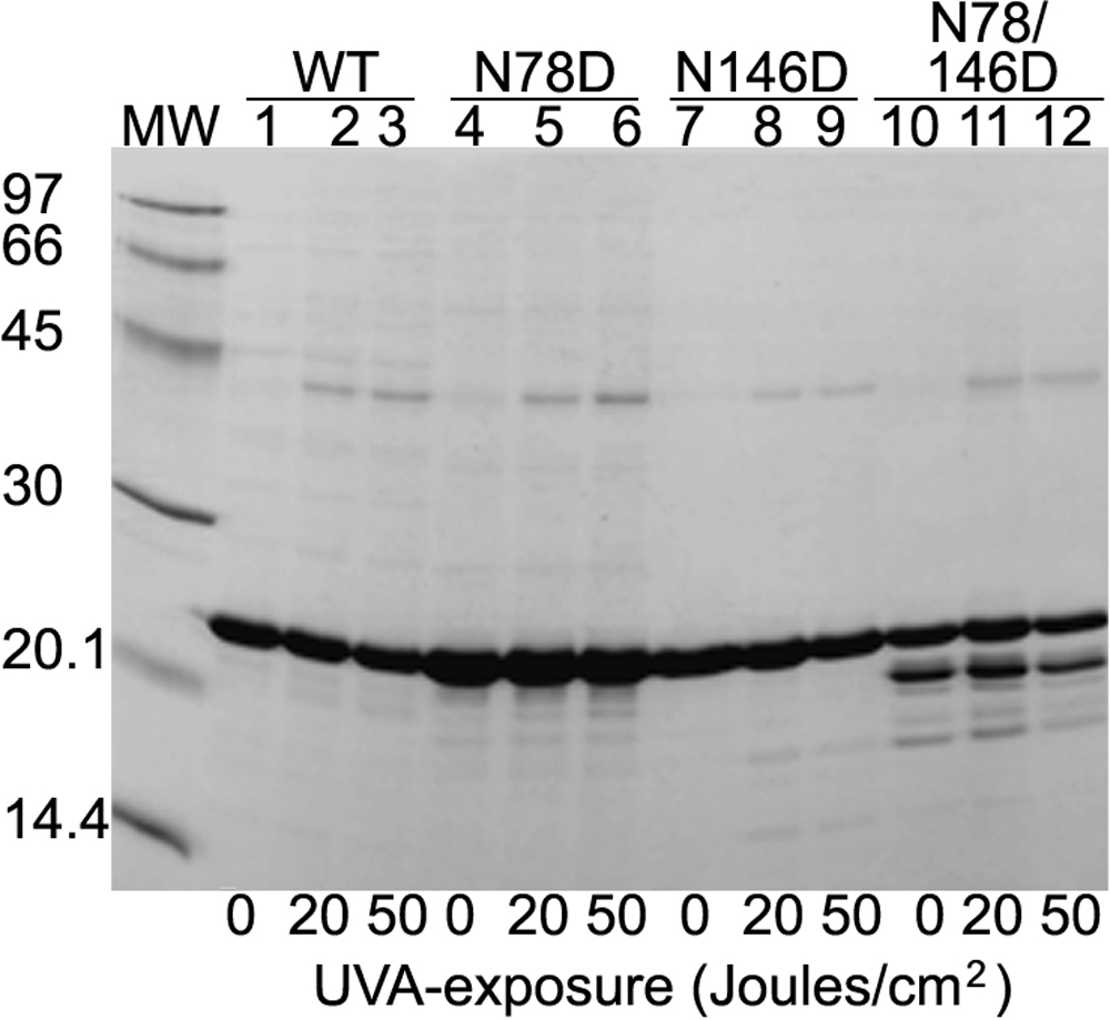
 Figure 1 of
Mafia, Mol Vis 2008; 14:234-248.
Figure 1 of
Mafia, Mol Vis 2008; 14:234-248.