Figure 6. Schematic representation of
light-induced PDE translocation on rod outer segment disc membrane.
Panel A shows schematic drawing of photoreceptor with
specialized rod outer segment domain containing stacks of discs
embedded with visual signaling proteins of rhodospin, transducin, and
phosphodiesterase (PDE). Abbreviations: rod outer segment (ROS); rod
inner segment (RIS). Panel B illustrates enlarged ROS disc
membranes with PDE molecules showing a hypothesized random distribution
of PDE molecules. Light induces a centripetal translocation of PDE away
from the rim-concentrated distribution in dark-adapted states.
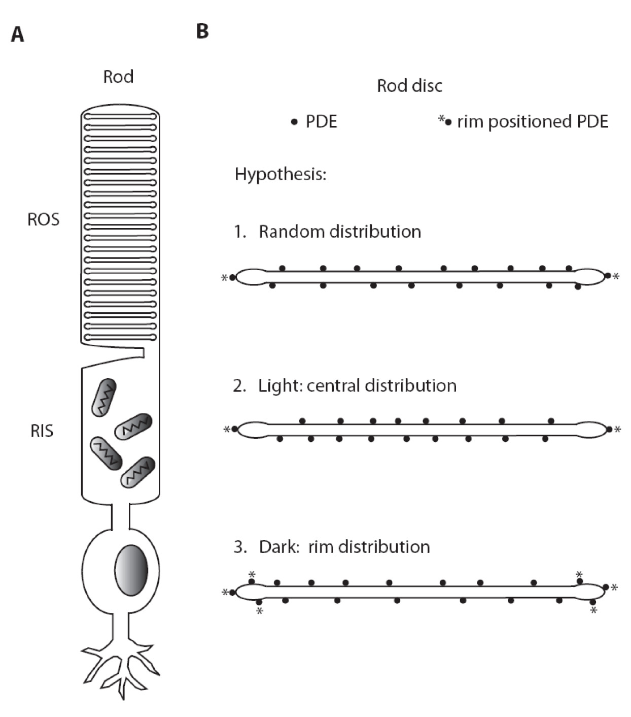
 Figure 6 of Chen, Mol Vis 2008; 14:2509-2517.