Figure 4. Effects of guanine nucleotides
on PDE localization. Rod outer segment (ROS) from dark-adapted rats
were isolated and permeabilized as described in Methods and
subsequently mixed with either 0.5 mM GTP, 0.5 mM GTPγS, or no
nucleotide. ROS suspensions were kept either in the dark or were
exposed to room light for 30 min, followed by fixation and preparation
for immunogold electron microscopy with PDEα antibody. The distances of
each gold particle from the nearest ROS plasma membrane were measured,
and the distribution profile of phosphodiesterase (PDE) was plotted.
Panels A and B represent PDE distribution in both
light- and dark-adapted conditions with guanosine triphosphates (GTP)
and GTPγS, respectively. Panel C represents the distribution of
PDE without additional nucleotides.
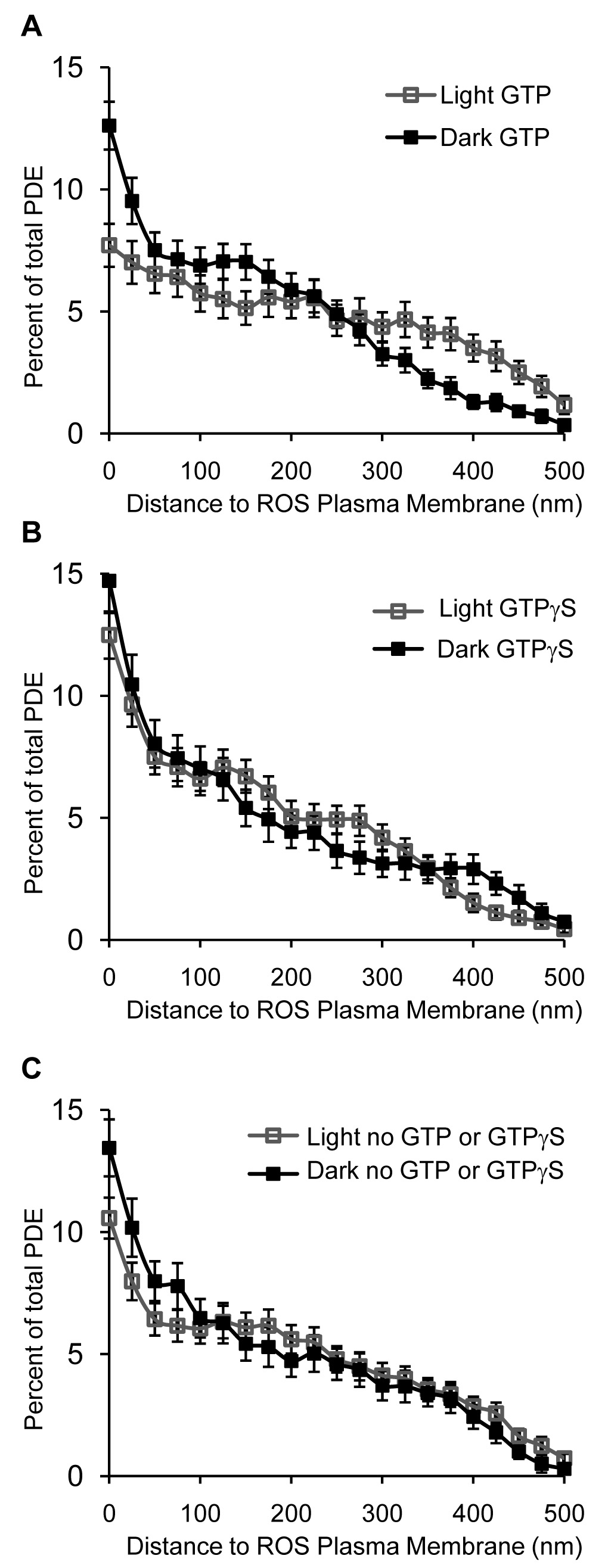
 Figure 4 of Chen, Mol Vis 2008; 14:2509-2517.