Figure 3. Time course of phosphodiesterase
translocation. Rod outer segment (ROS) isolated from dark-adapted rats
were exposed to room light (approximately 500 Lux) for various lengths
of time, followed by fixation and preparation for immunogold electron
microscopy. Percentage of total phosphodiesterase (PDE) adjacent to the
plasma membrane (within 25 nm) was measured and plotted (p≤0.002 for 0
versus 1 min).
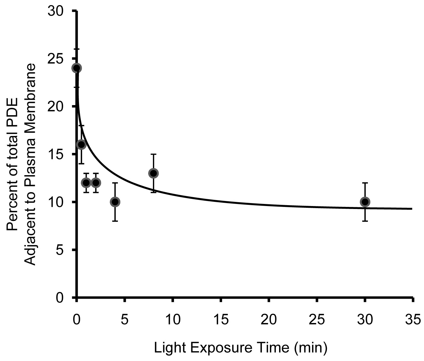
 Figure 3 of Chen, Mol Vis 2008; 14:2509-2517.