Figure 1. Photomicrograph pane showing
histological development of the anterior chamber angle. At F7w (A),
the angle structures are absent. At F10w and F15w (B and D),
the primitive anterior chamber angle was formed by densely packed
cells. At F18w and beyond (E), the cells at the iridocorneal
angle decreased in number and density. Between F15w and F27w (D–J),
the gradual appearance of immature trabecular beams with sparse
intertrabecular spaces and lined by spindle shaped trabecular meshwork
cells was noted. Note the anteriorly inserted ciliary body and
incompletely cleaved angle from F7w to F27w. Schlemm’s canal appeared
at F21w (G). A structurally mature Schlemm’s canal and a well
developed trabecular meshwork was observed at F27w (J) and
beyond, and a well developed trabecular meshwork was observed at P5m (K)
and in the adult eye (L). F, fetus; w, week; m, month; y, year;
C, Cornea; P, postnatal. The asterisk indicates the location of
Schlemm’s canal. An arrow indicates an iridocorneal angle. Double
arrows indicate the trabecular meshwork. Scale bar=50 μm.
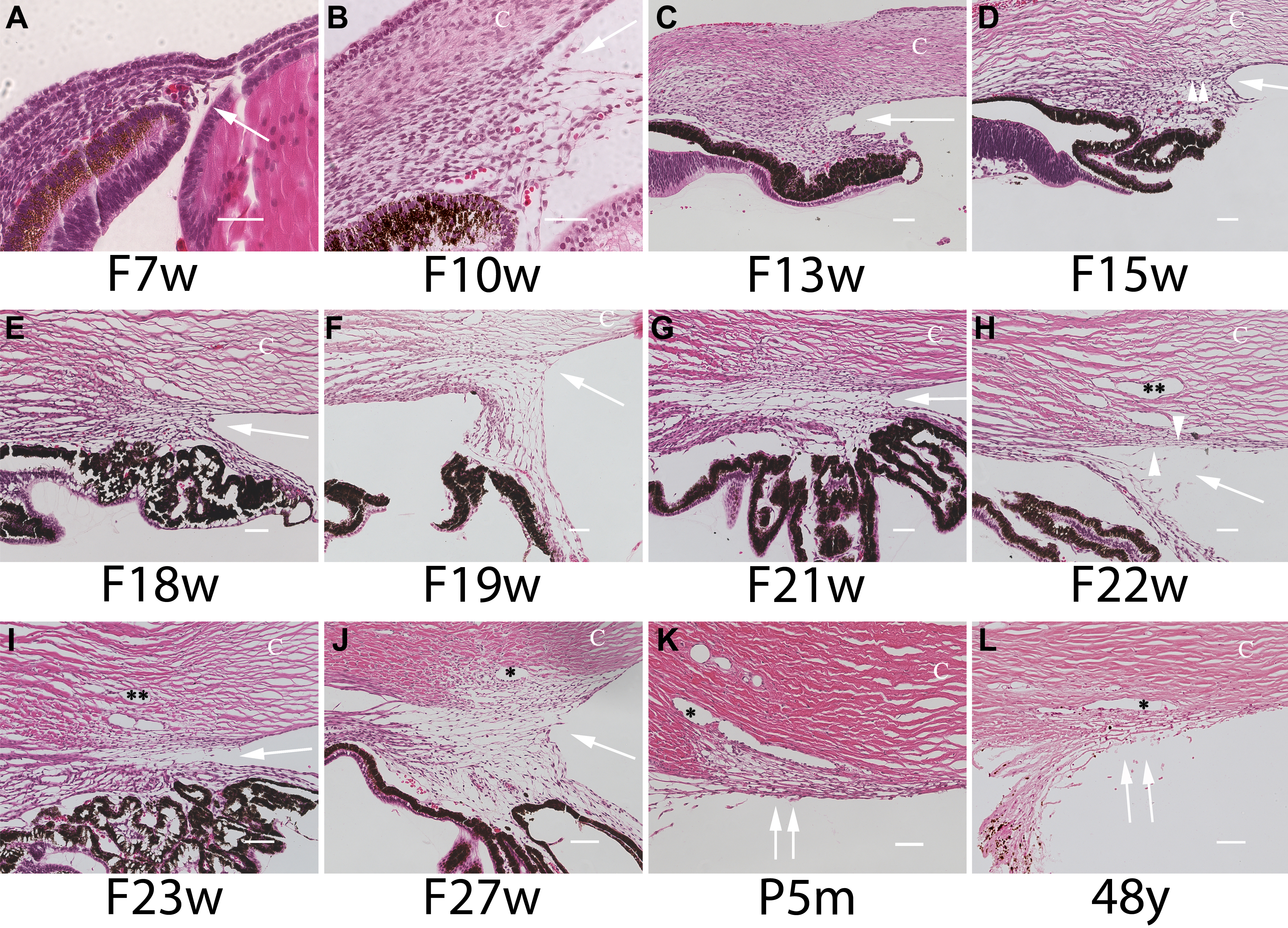
 Figure 1 of Meghpara, Mol Vis 2008; 14:2492-2498.