Figure 7. In isolated whole mount retina, GFP-labeled neurons exhibit spontaneous spikes. Merged fluorescence and infrared images of
a Tg(−12th:MmGFP) neuron and recording electrode in a whole mount zebrafish retina is shown in panel A. Scale bar equals 10 μm. Panel B displays spontaneous spikes recorded in a GFP-labeled cell. Loose-patch recordings were made using a voltage-clamp mode with
an electrode holding potential of 0 mV.
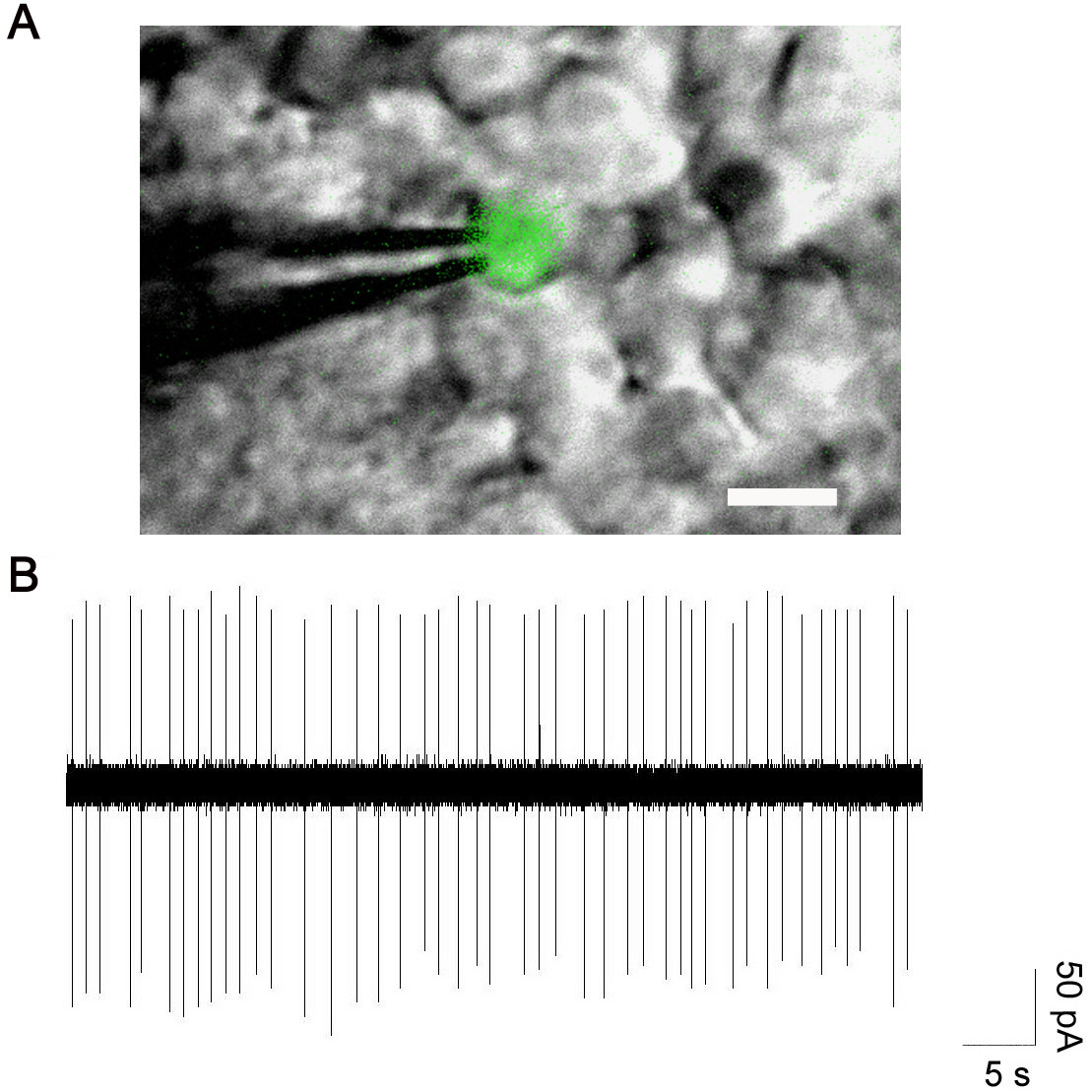
 Figure 7 of
Meng, Mol Vis 2008; 14:2475-2483.
Figure 7 of
Meng, Mol Vis 2008; 14:2475-2483.