Figure 4. Morphology of GFP-expressing cells in the retina. A: Localization of GFP-positive cells can be seen in this vertical retinal section. Most of the GFP-expressing cells were found
at the proximal cellular row of the inner nuclear layer. Abbreviations: ONL: outer nuclear layer (ONL); OPL: outer plexiform
layer (OPL); INL: inner nuclear layer; IPL: inner plexiform layer. B: This z-stack image shows the somata and processes of a single cell. C, GFP fluorescent fiber network in the inner plexiform layer. Scale bar equals 20 μm for A and C, and 10 μm for B.
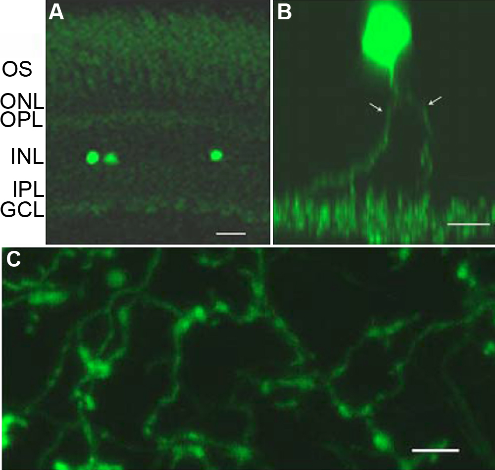
 Figure 4 of
Meng, Mol Vis 2008; 14:2475-2483.
Figure 4 of
Meng, Mol Vis 2008; 14:2475-2483.