Figure 3. Double immunostaining using
anti-GFP and anti-TH antibodies. Immunostaining experiments were
performed in vertical retinal sections (A) and the whole-mount
retina (B). GFP-IR, TH-IR, and GFP-IR/TH-IR are shown in green,
red and yellow, respectively. Quantification of double-staining in the
whole-mount retina is shown in C. Values represent the mean±SD
from 4 individual retinas. The cell density was calculated by dividing
the number of cells by the image area calculated by MetaMorph. Overall,
29±2% of GFP-labeled cells coexpressed TH. Scale bar equals10 μm for A
and 20 μm for B. Arrowhead in A points to a
GFP-IR/non-TH-IR cell. Arrowhead in B points to a
TH-IR/non-GFP-IR cell.
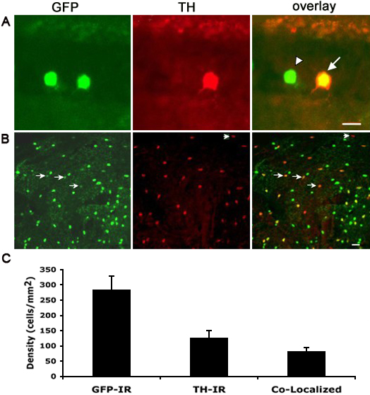
 Figure 3 of Meng, Mol Vis 2008; 14:2475-2483.