Figure 4. sCnx cells have increased
ubiquitylated inclusions. Wild-type (WT; A) and calnexin (sCnx;
B) cells were transfected with a His6-myc-tagged
ubiquitin expression plasmid and were stained with anti-myc antibody 24
h after transfection. Scale bar equals 10 μm. C: Cells were
scored for diffuse or punctate ubiquitin stain as exhibited in (A)
and (B). D: P23H rod opsin-green fluorescent protein
(GFP) and myc-ubiquitin double transfected cells were scored for
diffuse or punctate ubiquitin stain. Bar graphs represent an average of
three independent experiments. Error bars represent ±2 SEM. Statistical
analysis was performed using ANOVAR followed by posthoc tests. The
asterisk indicates p<0.05.
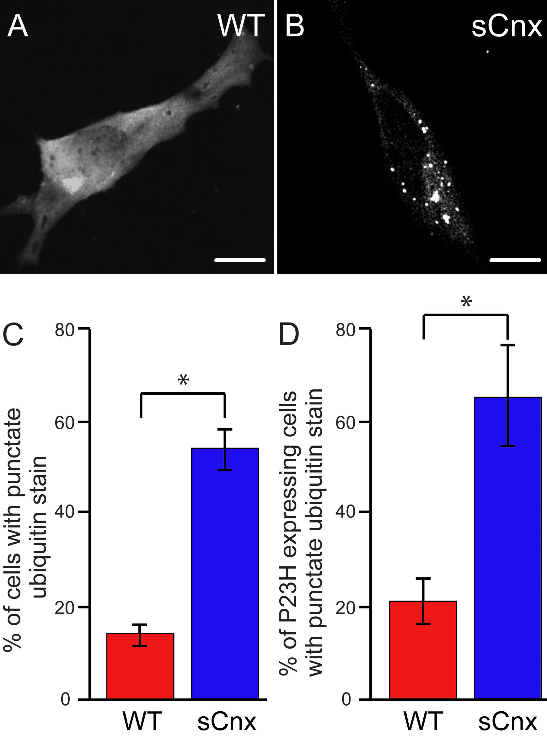
 Figure 4 of Kosmaoglou, Mol Vis 2008; 14:2466-2474.