Figure 1. Cnx expression in control and
sCnx cells. A: Localization of calnexin (Cnx) protein in
wild-type (WT) mouse embryonic fibroblasts (MEFs). B: Short Cnx
localization in sCnx cells. The truncated protein shows the same
staining pattern, as it still contains the N-terminal endoplasmic
reticulum (ER)-targeting sequence and the C-terminal ER retention motif
of full-length Cnx. Note the intensity of the sCnx staining was lower
and has been adjusted to reveal the pattern. Scale bar equals 10 μm. C:
Expression of ER resident chaperones Cnx and BiP in WT and sCnx cells.
Western blot revealed reduced expression level and increased
electrophoretic mobility of the truncated Cnx protein from sCnx cells
and similar BiP levels in both cell lines.
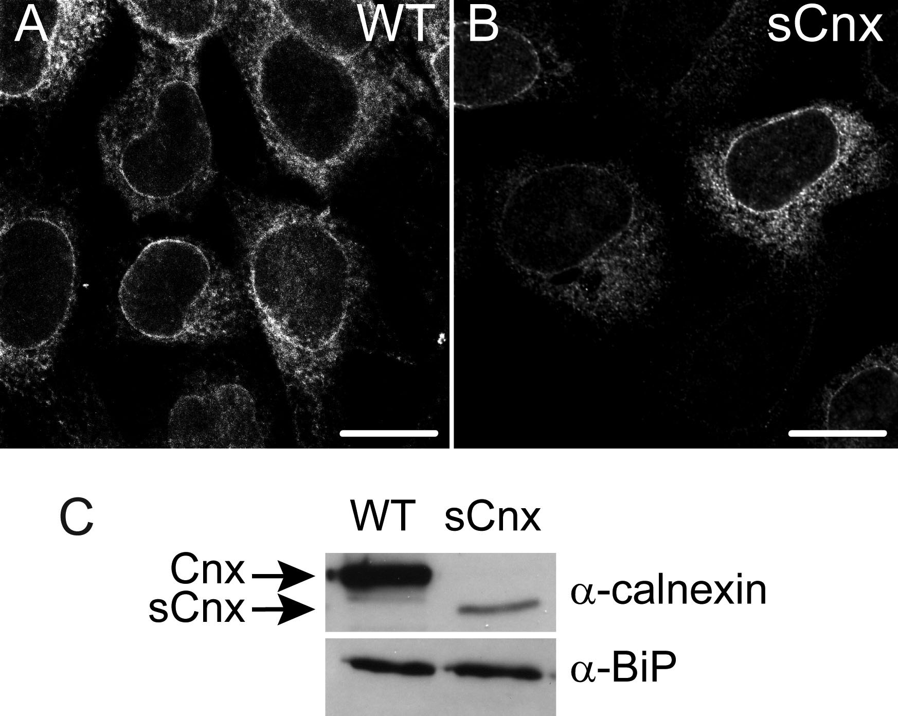
 Figure 1 of Kosmaoglou, Mol Vis 2008; 14:2466-2474.