Figure 3. CCR4 immunostaining of
conjunctiva and ciliary bodies in a model of endotoxin-induced uveitis
analyzed with a confocal microscope with 20X enlargement. Conjunctiva
was stained with CCR4 (in green) and propidium iodide for nuclei (in
red) in control rats (A) and of EIU rats 6 h (B) and 24 h
(C) after LPS injection. Staining of ciliary bodies was
performed in control rats (D) and in EIU rats at 6 h (E)
and 24 h (F). Immunostaining (in green) was positive in all
cases but comparable in the conjunctiva and the ciliary body.
Immunostaining did not show any clear differences between control rats
and rats with EIU.
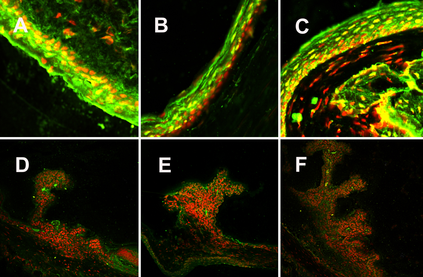
 Figure 3 of Trinh, Mol Vis 2008; 14:2428-2434.