Figure 2. Immunostaining in a model of endotoxin-induced uveitis analyzed with a confocal microscope with 20X enlargement. Conjunctivas
stained with anti-CCR5 antibody were revealed with secondary antibody, Alexa Fluor (in green), while nuclear chromatin was
stained with propidium iodide (in red) in control rats (A) and in EIU rats 6 h (B) and 24 h (C) after LPS injection. Ciliary bodies were stained with the same antibodies in control rats (D) and in EIU rats 6 h (E) and 24 h (F) after LPS injection. CCR5 expression (in green) was higher in the conjunctiva and ciliary bodies of the rats with EIU than
of the controls.
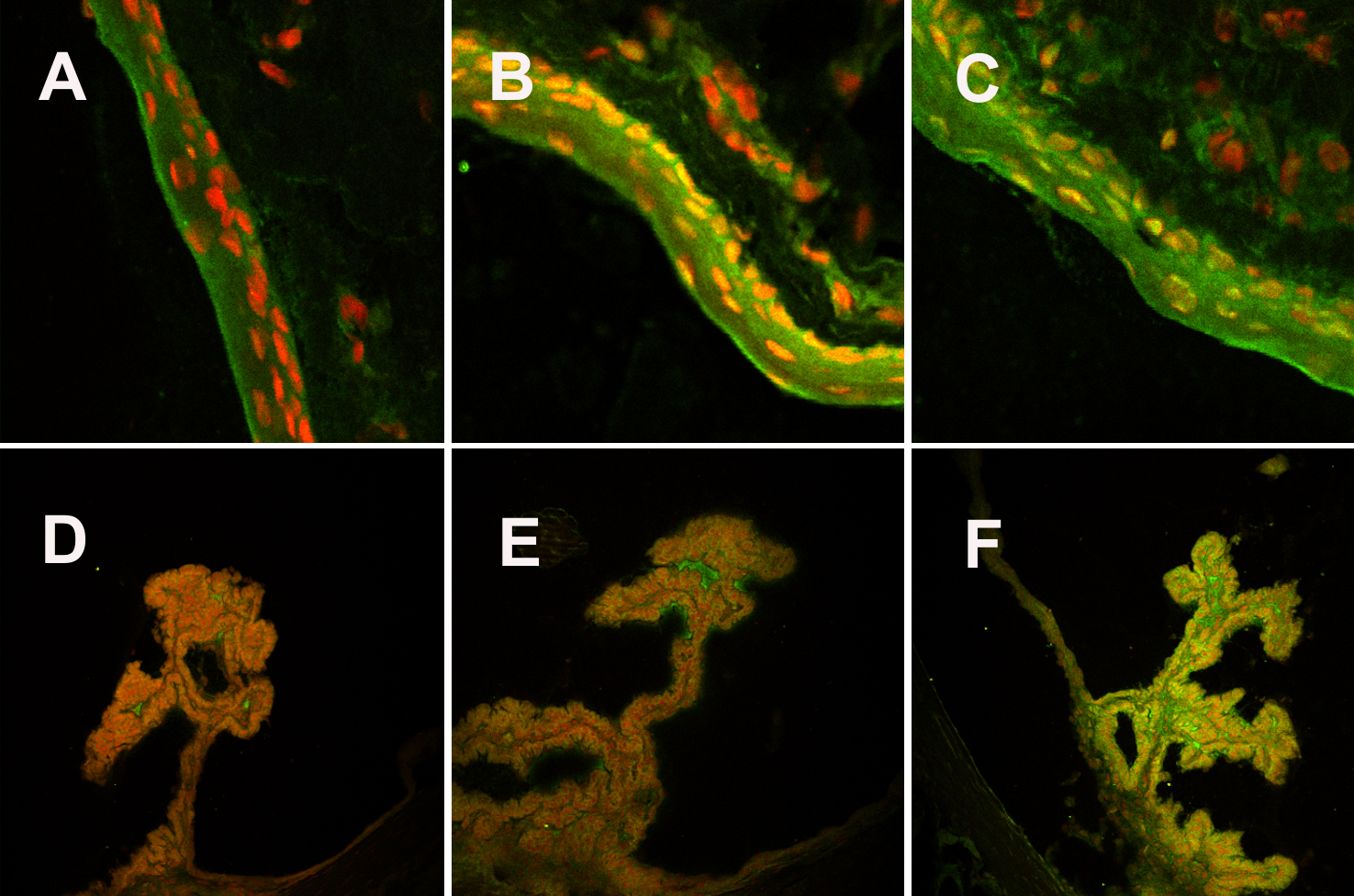
 Figure 2 of
Trinh, Mol Vis 2008; 14:2428-2434.
Figure 2 of
Trinh, Mol Vis 2008; 14:2428-2434.