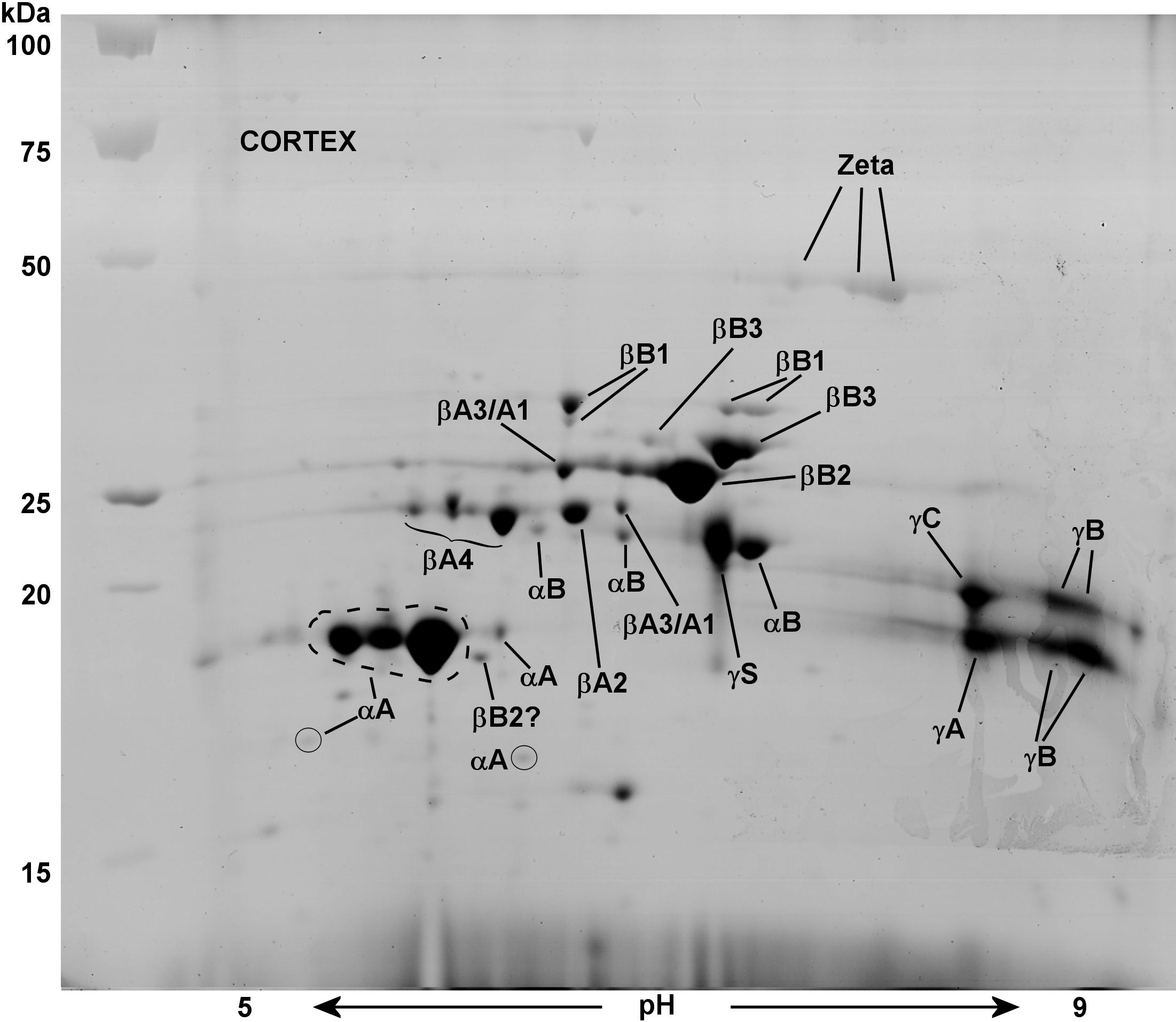
Figure 4. 2-D Electrophoresis map showing
identities of lens cortical water-soluble (WS) proteins of a
2.5-month-old guinea pig. The major proteins of the lens cortex were
identified by MALDI mass spectrometry. The ratio of cortical
αA-crystallin to αB-crystallin was 6.5:1 as quantified by image
analysis software,
Image J.
All β-crystallins and γA-C-crystallin were detected, but no protein
signatures were detected for γD-, γE-, and γF-crystallin. The gel
contains 47 spots, 37 of which have been identified, with some proteins
as intact or truncated crystallins. Proteins were stained with
Coomassie Blue G-250. The symbol ? indicates presumptive protein
identification.
 Figure 4 of Simpanya, Mol Vis 2008; 14:2413-2427.