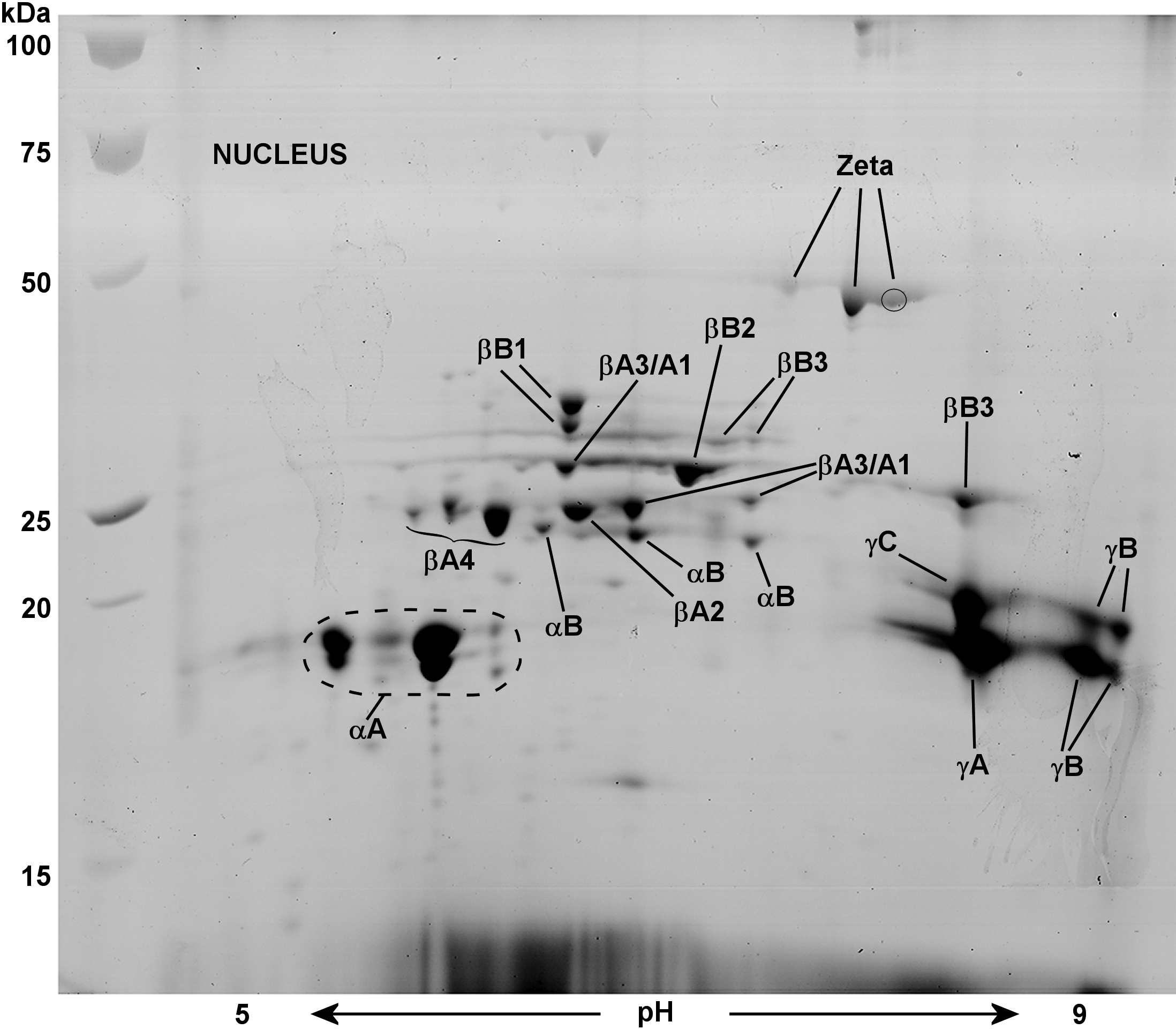
Figure 3. 2-D Electrophoresis map showing
identities of lens nuclear water-soluble proteins of a 2.5-month-old
guinea pig. The major proteins of the lens nucleus were identified by
matrix assisted laser desorption (MALDI) mass spectrometry.
αA-crystallin was far more abundant than αB-crystallin (ratio of 8:1)
as quantified by image analysis software,
Image J. All β-crystallins
(βA1-, βA2-, βA3-, βA4-, βB1-, βB2-, and βB3-crystallin) and γA-, γB-,
and γC-crystallin were detected on 2-DE gels, but no protein signatures
were found for γD-, γE-, or γF-crystallin. The gel contains 53 spots,
44 of which have been identified as various intact or truncated
crystallins. Proteins were stained with Coomassie Blue G-250.
 Figure 3 of Simpanya, Mol Vis 2008; 14:2413-2427.