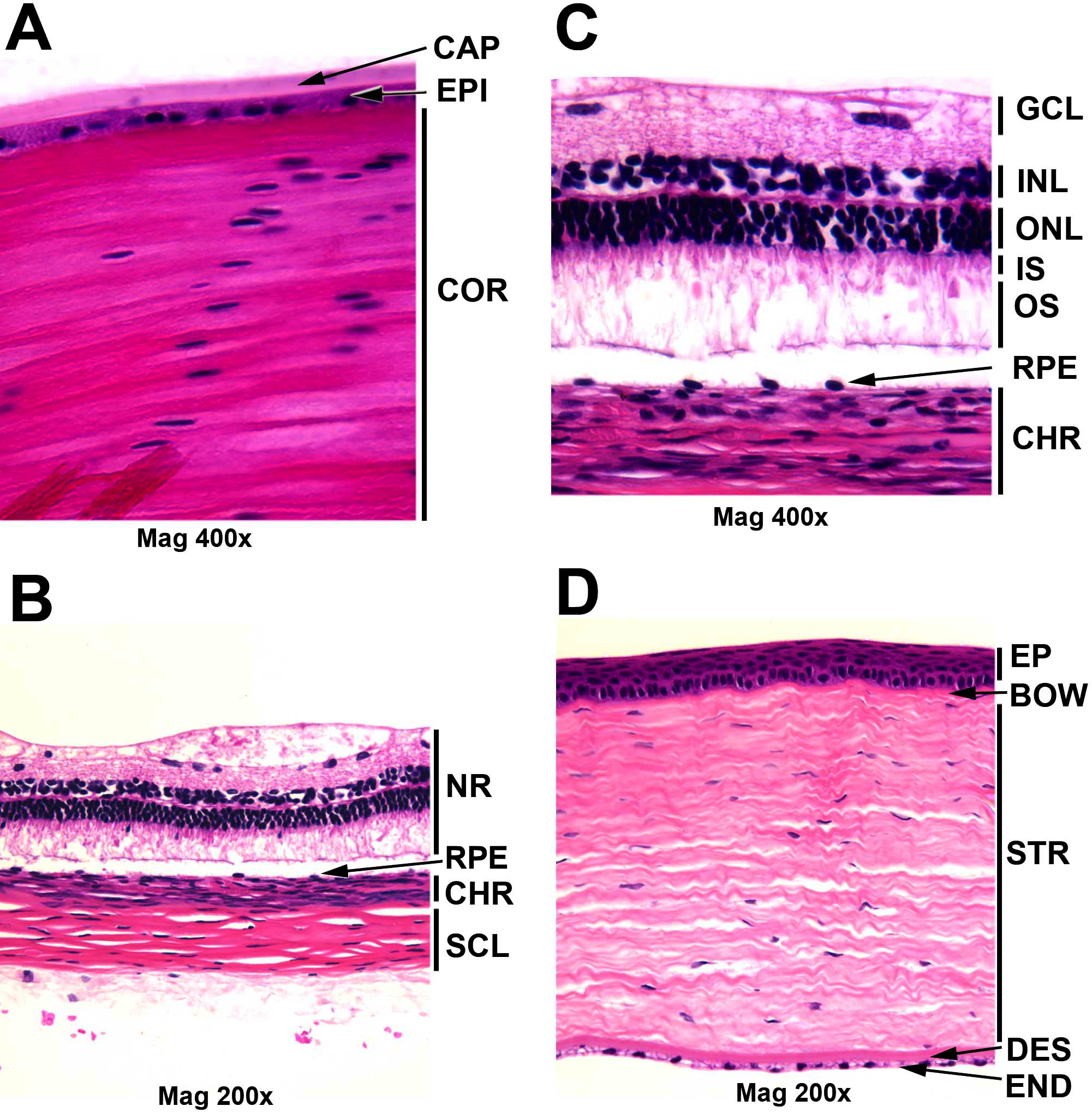
Figure 1. Morphology of guinea pig eye
tissues for the Hartley strain used for the NEIBank library, stained
with hematoxylin and eosin reagent. A: Lens capsule, epithelium
and cortex in the bow region: capsule (CAP), epithelium (EPI), and
cortical fiber cells (COR). The guinea pig lens is similar to human and
mouse. B:
Neural retina (NR), retinal pigment epithelium (RPE), choroid (CHR) and
sclera (SCL). C: Retinal layer and choroid: ganglion cell layer
(GCL), inner nuclear layer (INL), outer nuclear layer (ONL), inner
segment (IS), outer segment (OS), retinal pigment epithelium (RPE), and
choroid (CHR). The guinea pig retina is 4–5 nuclei thick, similar to
the human retina. D: Cornea: corneal epithelium (EP), Bowman’s
membrane (BOW), Stroma (STR), Descemet’s membrane (DES) and endothelium
(END). The guinea pig cornea is similar to human, while the mouse has a
thinner stroma.

 Figure 1 of Simpanya, Mol Vis 2008; 14:2413-2427.
  Figure 1 of Simpanya, Mol Vis 2008; 14:2413-2427.