Figure 5. Two-dimensional electrophoresis photography of regenerated lens. A-C: Patterns of protein spots in regenerated lenses (two weeks, four weeks, and 16 weeks). D-F: Patterns of protein spots of natural lenses (two weeks, four weeks, and 16 weeks). The protein patterns are very similar
among all stages of regeneration (two weeks, four weeks, and 16 weeks) and shared a high degree of analogy with those of 16-week-old
natural lenses (F). However, these patterns were significantly different from those of two-week-old or four-week-old natural lenses (D,E).
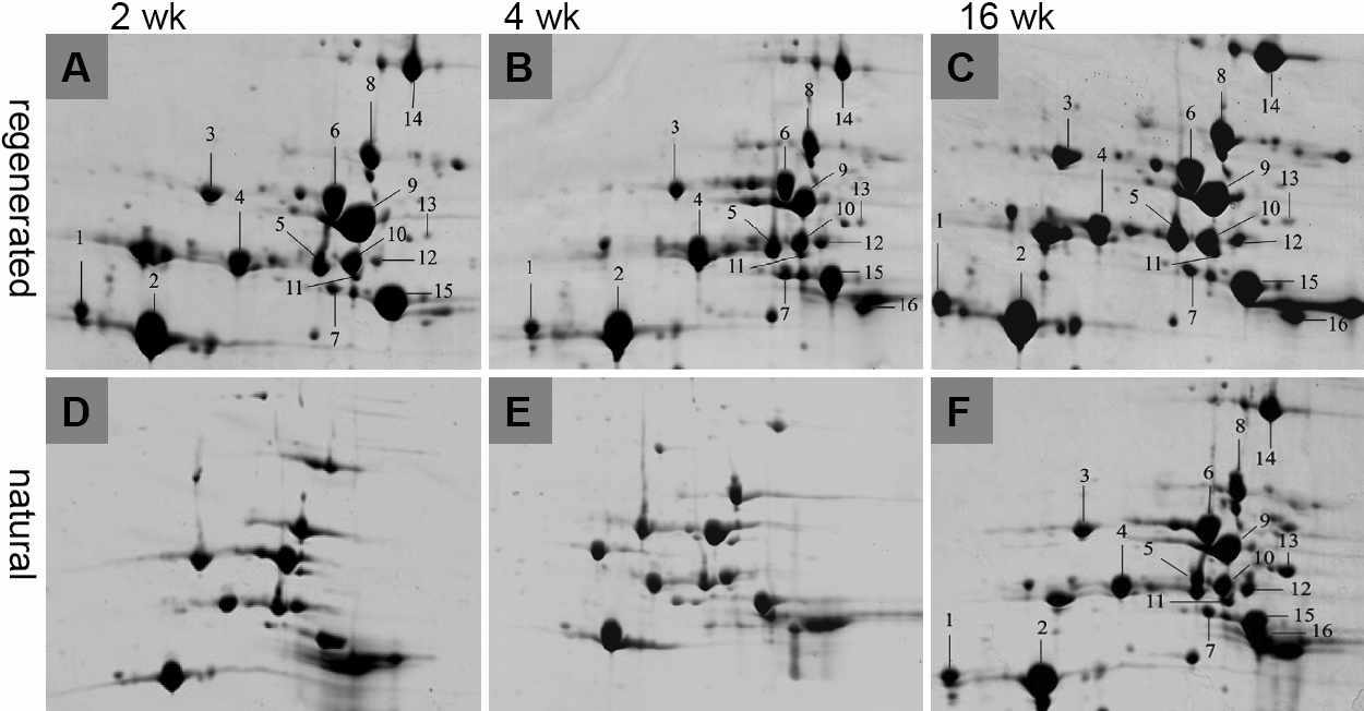
 Figure 5 of
Liu, Mol Vis 2008; 14:2404-2412.
Figure 5 of
Liu, Mol Vis 2008; 14:2404-2412.