Figure 4. Representative transmission electron micrograph of epithelial cells in a regenerated lens. Morphological changes are seen
both at the peripheral equatorial (A) and central (B) areas, including overly dense and indented nuclei, edematous mitochondria, and an expanding endoplasmic reticulum when compared
to natural lenses (C,D). Magnification is8,000X).
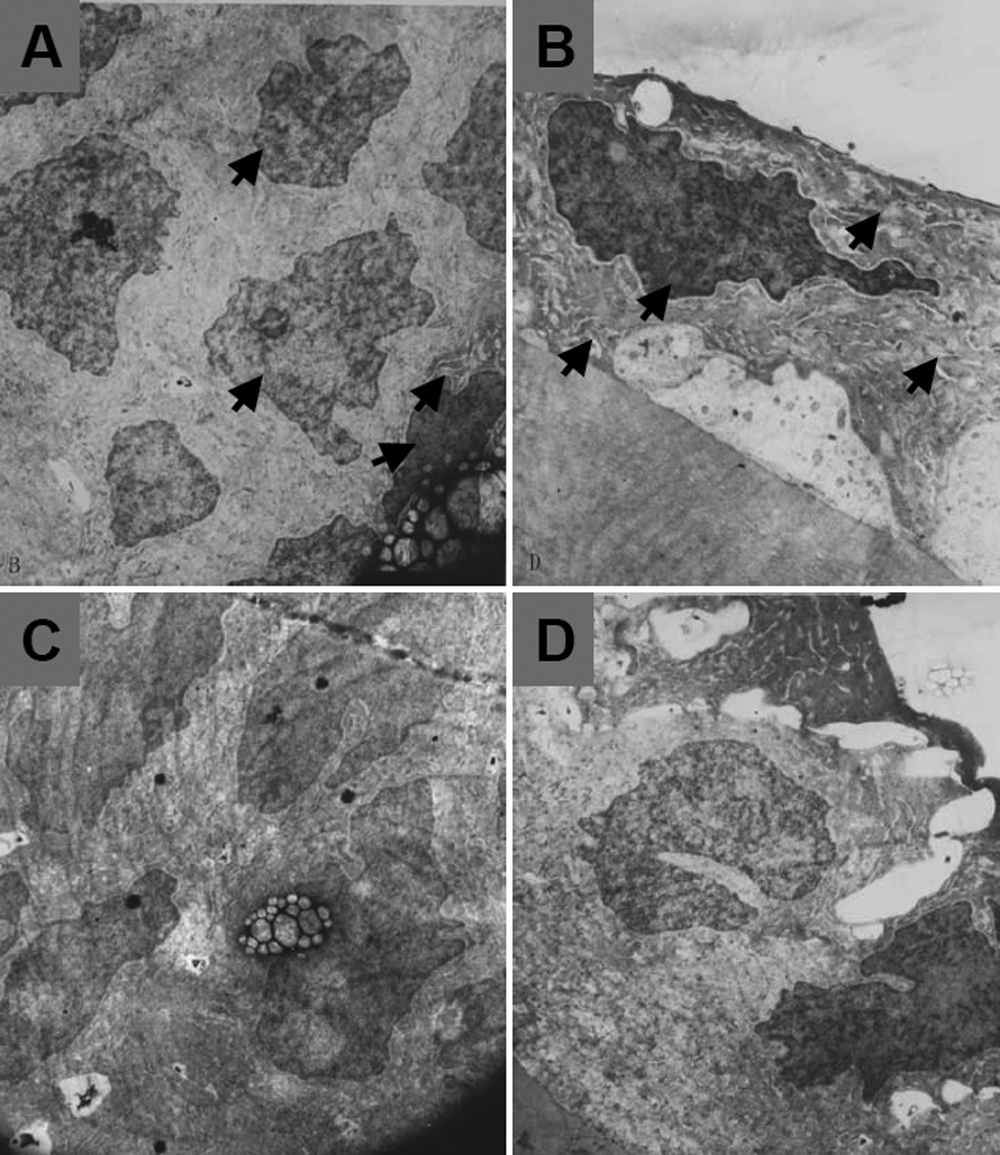
 Figure 4 of
Liu, Mol Vis 2008; 14:2404-2412.
Figure 4 of
Liu, Mol Vis 2008; 14:2404-2412.