Figure 3. Lgl1 mRNA distribution in
control retina and protein localizations in adult retina and during
development from control and Trp1/Tag mice using immunohistochemistry. A
and B show Lgl1 mRNA localization whereas C-N
show Lgl1 protein distribution in the adult mouse retina from the
control mouse (C-E) and Trp1/Tag mouse model (F-N)
at P15 (F), P20 (G,H), P25 (I,J),
P30 (K,L) and three months (M,N). A
and C show the negative controls for B and D,
respectively. In the retina, Lgl1 mRNA and protein were found
in the ganglion cell layer (GCL), the inner nuclear layer (INL), the
outer nuclear layer (ONL), the photoreceptor inner segments (IS), the
external limiting membrane (elm), the RPE, and the melanocytes and
vascular cells in the choroid (Ch; B). No mRNA staining was
found in the inner plexiform layer (IPL) or in the outer plexiform
layer (OPL; B), but a weak trace of protein was observed (D).
Lgl1 labeling in the retinal nuclear layers seemed to be exclusively
membranous (D,E). Lgl1 protein distribution in the
retinas from the Trp1/Tag mouse model changed during tumoral
development. In the INL and the RPE, Lgl1 immunolabeling appeared to be
both membranous and cytoplasmic compared to the control retina (F,G,I,K).
Lgl1 protein also appeared to be reduced in the INL, the OPL, and the
elm from P25 (I,K). The tumor cells (TC) also displayed
staining for Lgl1 protein during tumoral development in Trp1/Tag mice (H,J,L,N).
However, very weak Lgl1 staining was detected in tumor cells at P30 (L).
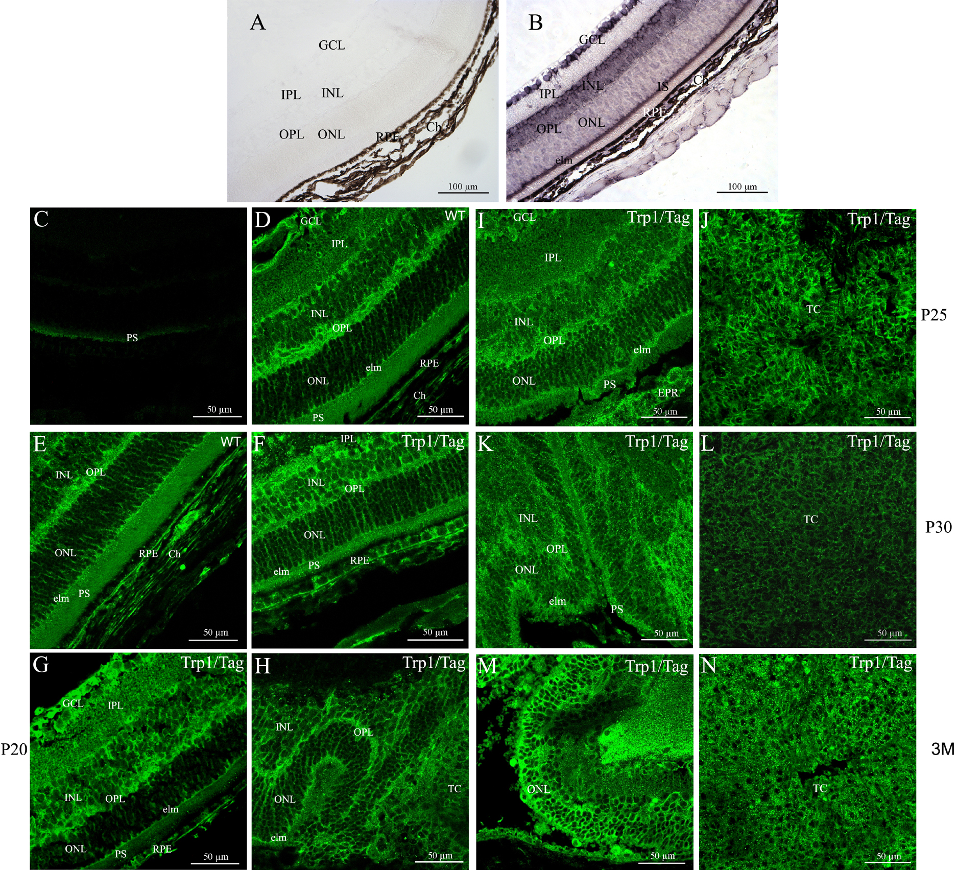
 Figure 3 of Vieira, Mol Vis 2008; 14:2390-2403.