Figure 2. Scrib mRNA distribution
in control retina and protein localizations in adult retina and during
development from control and Trp1/Tag mice using immunohistochemistry. A
and B show Scrib mRNA localization whereas C-P
show Scrib protein distribution in adult mouse retina from the control
mouse (C-E) and the Trp1/Tag mouse model (F-P)
at P15 (F), P20 (G,H), P25 (I-K), P30 (L-N),
and three months (O,P). A and C show the
negative controls for B and D, respectively. In the
retina, the Scrib mRNA and protein were observed in the
ganglion cell layer (GCL), the inner nuclear layer (INL), the outer
nuclear layer (ONL), and the outer plexiform layer (OPL). No mRNA
expression was detected in the inner plexiform layer, but its protein
was detected (in the IPL). Very strong levels of expression were
detected in the photoreceptor inner segments (IS) and the external
limiting membrane (elm). We also detected Scrib in the retinal pigment
epithelium (RPE), choroidal melanocytes, and choroidal vascular cells
(Ch). The neural retina continued staining positive for the Scrib
protein in ocular tumor development in Trp1/Tag mice (F-P).
However, from P15, strong staining was lost in the OPL (F,G,I,L).
Scrib immunolabeling in the nuclear layers appeared to be more diffuse
rather than restricted to cell membranes as observed in the control
retina (F,G,I,L). The thickening of the
pigmentary epithelium (RPE) was also positive for Scrib, which was
predominantly localized to the cell membrane but also present in the
cytoplasm (F). In addition, low levels of Scrib seemed to be
present in the uvea (Ch; M). The intensity of the Scrib protein
labeling in tumoral cells was markedly reduced and no longer restricted
to cell membranes as described for P25. Scrib protein was detectable in
tumor cells from P20 (H,K). However, at P30 and three
months, no significant Scrib immunolabeling could be detected in
tumoral cells (N,P).
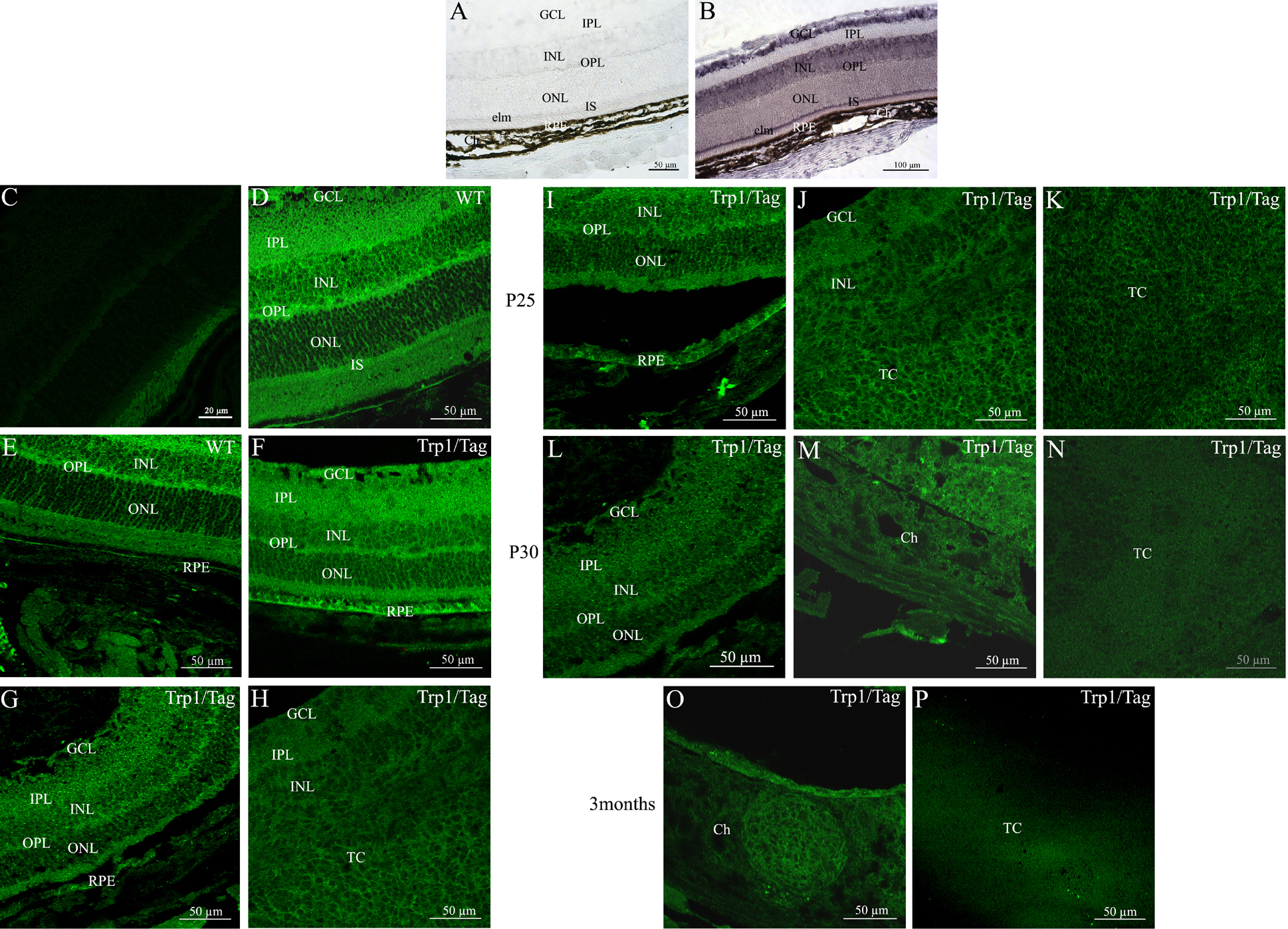
 Figure 2 of Vieira, Mol Vis 2008; 14:2390-2403.