Figure 1. Dlg1 mRNA distribution in
the control retina and protein localizations in the adult retina and
during development from control and Trp1/Tag mice using
immunohistochemistry. A and B show Dlg1 mRNA
localization whereas C-K show the Dlg1 protein
distribution in the adult mouse retina from the control mouse (C
and D) and from the Trp1/Tag mouse model (E-K) at
P15 (E), P20 and P25 (F-H), P30, and three months (I-K).
A and C show the negative controls for B and D,
respectively. Dlg1 mRNA and protein distributions were widely expressed
in the ganglion cell layer (GCL), the inner nuclear layer (INL), the
outer nuclar layer (ONL), and the photoreceptor inner segments (IS) of
the control mouse retina. Very strong staining was detected in the
outer plexiform layer (OPL) and the external limiting membrane (elm).
However, faint staining of Dlg1 mRNA was found in the inner
plexiform layer whereas its protein was strongly detected. Futhermore,
in the inner nuclear layer, Dlg1 labeling was stronger at
either side of the layer than in its center (indicated by an asterisk; D).
Retina from the transgenic Trp1/Tag mouse model of ocular tumor
continued to express Dlg1 protein (E-K). However, changes
in the distribution of Dlg1 protein was observed from P20. In the
central region adjacent to the mass of tumoral cells, Dlg1
immunolabeling in photoreceptor segments (IS) seemed to be reduced (F,G,I).
A lower intensity of Dlg1 immunolabeling was shown in the OPL compared
to the control retina (F,G,I). We also observed
that immunolabeling in the INL appeared to be more diffuse in
transgenics than in controls, possibly reflecting more labeling at the
cortical regions than at membranous regions (F,I). In
addition, from P30, the RPE and choroid (Ch) no longer stained positive
for Dlg1 protein (J) in the posterior pole. The tumor cells (TC)
had very faint staining for Dlg1 protein at these stages (H,K).
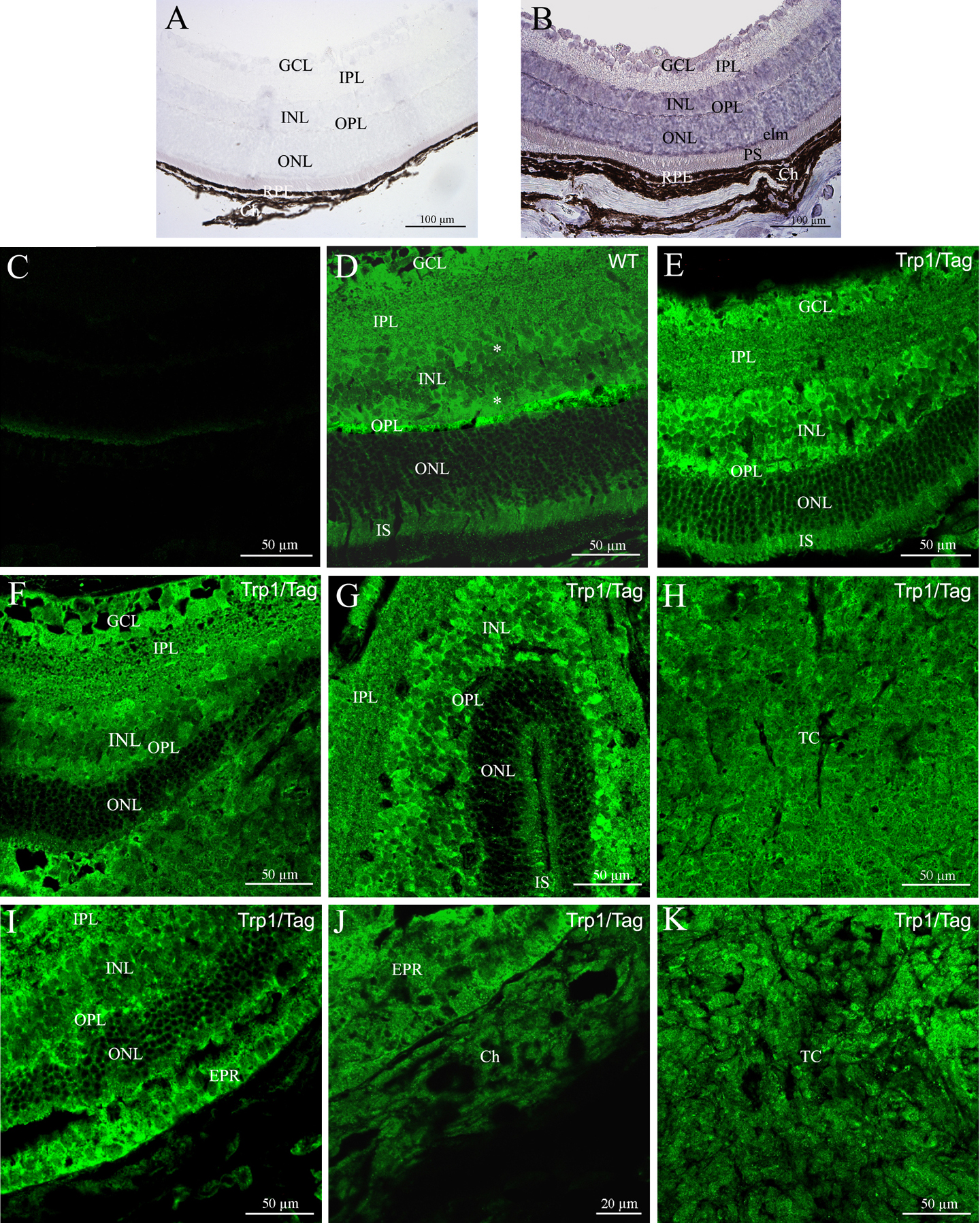
 Figure 1 of Vieira, Mol Vis 2008; 14:2390-2403.