Figure 2. Distribution of BMP-2 in human sclera fibroblasts in vitro using indirect immunofluorescence. FITC marked the secondary antibody
(green; A), and PI dyed the nucleus (red; B). The first (A) and second images (B) are combined to form the third image shown (C). BMP-2 is localized in the cytoplasm and weakly in the nucleus of HSF (A-C). Magnification: 400X.
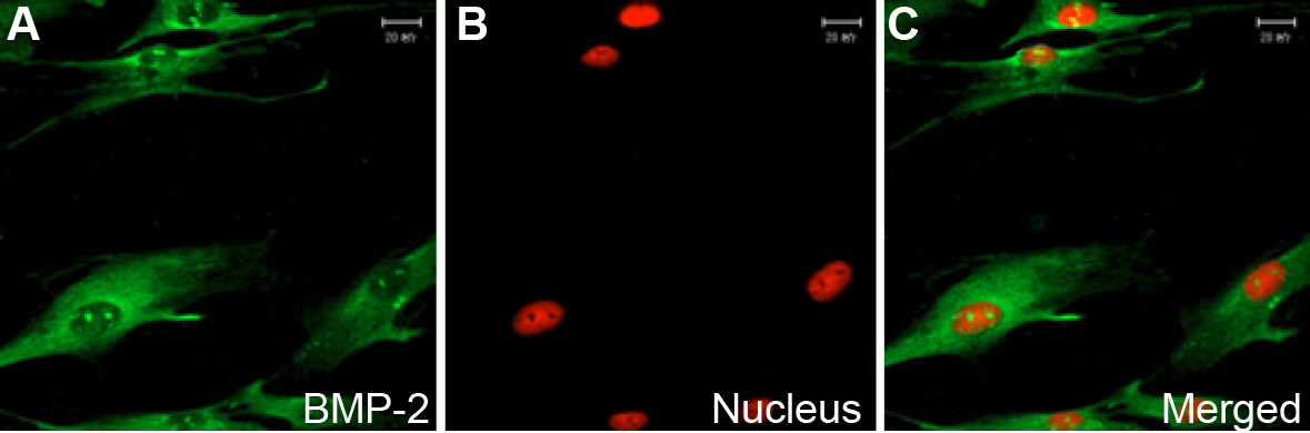
 Figure 2 of
Hu, Mol Vis 2008; 14:2373-2380.
Figure 2 of
Hu, Mol Vis 2008; 14:2373-2380.